Silfverskiold Test Interpretation
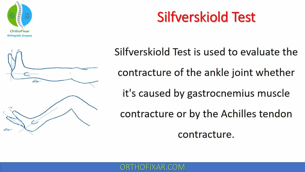
Silfverskiold Test is used to evaluate the contracture of the ankle joint whether it’s caused by gastrocnemius muscle contracture or by the Achilles tendon contracture. It measures the dorsiflexion of the foot at the ankle joint with knee extended and flexed to 90 degrees.
This test was first described by Nils Silfverskiöl in 1923.
How do you perform the Silfverskiold Test?
The Silfverskiöld Test is commonly used to identify isolated gastrocnemius contracture associated with several foot and ankle pathologies. Here’s how you perform it:
- Position the patient lying prone (face down) on the examination table or bed with the affected leg extended.
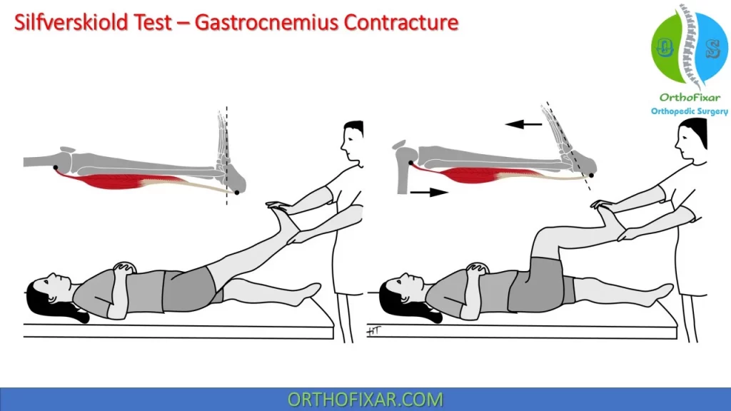
- The examiner dorsiflexes the foot while the knee in full extension and measures the ankle dorsiflexion range of motion.
- Then the test is repeated with knee flexed at 90 degrees and the ankle dorsiflexion range of motion is measured.
- The test can be done with the patient is sitting.
See Also: Ankle Range of Motion
See Also: Thomas Test
What does a positive Silfverskiold Test mean?
Ankle movements are influenced by the position of the knee because gastrocnemius crosses both joints.
Gastrocnemius Tightness: If the dorsiflexion increases significantly with the knee flexed, it suggests that the gastrocnemius muscle (which crosses the knee and ankle joints) is tight. This is because bending the knee relaxes the gastrocnemius muscle, allowing greater ankle dorsiflexion.
See Also: Gastrocnemius Muscle Anatomy
Achilles Tendon Contracture: If there is no significant change in the dorsiflexion angle with the knee bent, it suggests that the restriction is due to stiffness in the Achilles tendon or the soleus muscle, which does not cross the knee.
In all patient with ankle joint contracture, one should assess the spine for deformity and range of motion.
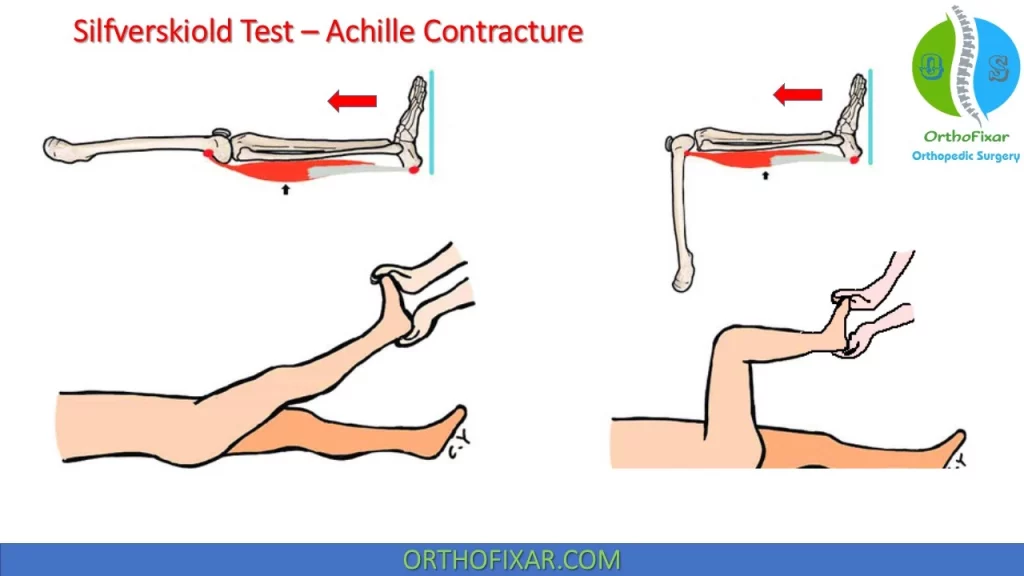
Because the gastrocnemius originates from above the knee, it tightens when the knee is extended and relaxes when the knee is flexed. If the loss of dorsiflexion is because of gastrocnemius tightness alone, then there will be reduced dorsiflexion with the knee extended. If there is no change of ankle dorsiflexion with knee flexion or extension, then the contracture is in both gastrocnemius and soleus.
Accuracy
The Silfverskiöld test had poor inter- and intrarater reliability.
Reverse Silfverskiold test
Reverse Silfverskiold test is not very popular.
With knee in full extension (ankle dorsiflexion here is solely restricted by tendo-Achilles) measure the range of dorsiflexion at ankle (more on injured side compared to the normal side).
Notes
Do not use the plantar medial border as the presence of supination or pronation deformity of the forefoot will give false recording and values if missed.
Few authors suggest that the Silfverskiold test should be done with the foot held in inversion while assessing the foot and ankle. The inversion locks the foot and thereby eliminates instability of the hindfoot and midfoot joints.
Flatfoot presents a special challenge when assessing for contracture of the heel cord. The reason is that both the ankle joint and subtalar joint dorsiflex and plantarflex. To isolate ankle joint dorsiflexion, the subtalar joint is anatomically aligned or locked using inversion to prevent subtalar dorsiflexion. The cavus foot presents a challenge to assess the heel cord contracture. Assessment of ankle equinus is done by isolating the hindfoot. The forefoot can be obscured from vision with the hand so that only the hindfoot is visualized.
Related Anatomy
Gastrocnemius Muscle Anatomy
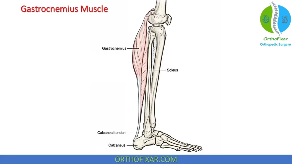
The gastrocnemius originates from above the knee by two heads, each head connected to a femoral condyle and to the joint capsule. Approximately halfway down the leg, the gastrocnemius muscles merge to form an aponeurosis. As the aponeurosis gradually contracts, it accepts the tendon of the soleus, a flat broad muscle deep to the gastrocnemius. The aponeurosis and the soleus tendon end in a flat tendon, called the Achilles tendon, which attaches to the posterior aspect of the calcaneus. The two heads of the gastrocnemius and the soleus are collectively known as the triceps surae.
Although the primary function of the gastrocnemius– soleus complex is to plantar flex the ankle and to supinate the subtalar joint, the gastrocnemius also functions to flex or extend the knee, depending on whether the lower extremity is weightbearing or not.
It has been proposed that weakness of the gastrocnemius may cause knee hyperextension. In addition, it has been theorized that the gastrocnemius acts as an antagonist to the ACL, exerting an anteriorly directed pull on the tibia throughout the range of knee flexion–extension motion, particularly when the knee is near extension.
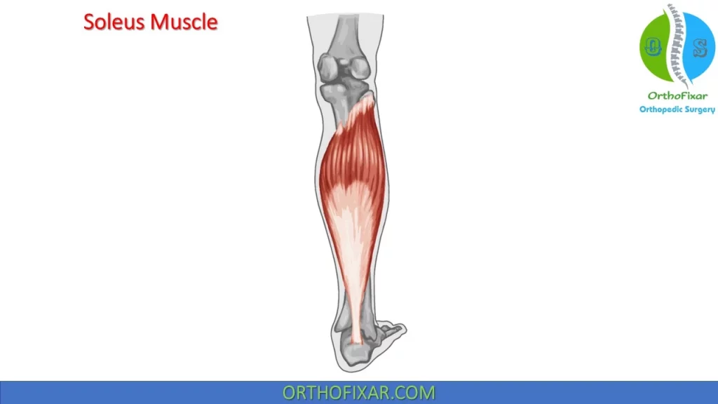
Soleus Muscle
The Soleus Muscle arises from the head of fibula, proximal third of shaft, soleal line and midshaft of posterior tibia. It inserts onto Posterior surface of calcaneus through Achilles tendon. It’s innervated by the tibial nerve.
See Also: Ankle Anatomy
Achilles Tendon
The Achilles tendon is the thickest, strongest tendon in the body. As the Achilles tendon comes off the posterior calf muscles, it courses distally to attach approximately three-quarters of an inch below the superior portion of the os calcis, on the medial aspect of the calcaneus.
References
- Symeonidis P. The Silfverskiöld Test. Foot Ankle Int. 2014 Aug;35(8):838. doi: 10.1177/1071100714535202. Epub 2014 Jul 30. PMID: 25080357.
- Baumbach SF, Brumann M, Binder J, Mutschler W, Regauer M, Polzer H. The influence of knee position on ankle dorsiflexion – a biometric study. BMC Musculoskelet Disord. 2014 Jul 23;15:246. doi: 10.1186/1471-2474-15-246. PMID: 25053374; PMCID: PMC4118219.
- DiGiovanni CW, Kuo R, Tejwani N, Price R, Hansen ST Jr, Cziernecki J, Sangeorzan BJ. Isolated gastrocnemius tightness. J Bone Joint Surg Am. 2002 Jun;84(6):962-70. doi: 10.2106/00004623-200206000-00010. PMID: 12063330.
- Fleiss DJ. Diagnostic tests for gastrocnemius tightness. J Bone Joint Surg Am. 2003 Apr;85(4):760; author reply 760. doi: 10.2106/00004623-200304000-00027. PMID: 12672856.
- Molund M, Husebye EE, Nilsen F, Hellesnes J, Berdal G, Hvaal KH. Validation of a New Device for Measuring Isolated Gastrocnemius Contracture and Evaluation of the Reliability of the Silfverskiöld Test. Foot Ankle Int. 2018 Aug;39(8):960-965. doi: 10.1177/1071100718770386. Epub 2018 Apr 20. PMID: 29676167.
- Clinical Assessment and Examination in Orthopedics, 2nd Edition Book.
- Millers Review of Orthopaedics -7th Edition Book.
- Lifetime product updates
- Install on one device
- Lifetime product support
- Lifetime product updates
- Install on one device
- Lifetime product support
- Lifetime product updates
- Install on one device
- Lifetime product support
- Lifetime product updates
- Install on one device
- Lifetime product support