Posterior Drawer Test of the Ankle

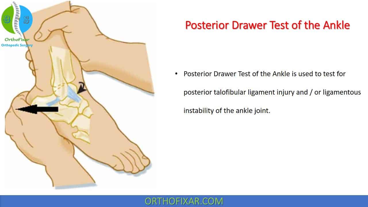
Posterior Drawer Test of the Ankle is used to test for posterior talofibular ligament injury and / or ligamentous instability of the ankle joint.
Ankle Posterior Drawer Test was first described by Frost and Hanson in 1977.
See Also: Ankle Anatomy
How Posterior Drawer Test of the Ankle is Performed?
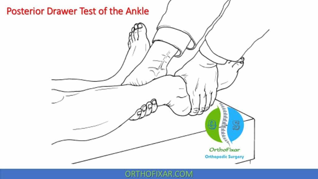
Posterior Drawer Test of the Ankle is performed with the patient lies supine with the knee slightly flexed to neutralize the pull of the gastrocnemius muscle.
With the ankle joint held at 10 to 15° of plantar flexion, the examiner grasps around the heel with one hand and stabilizes the tibia from the anterior side with the other.
After asking the patient to relax the muscles, the examiner moves the foot posteriorly at the ankle
joint while continuing to hold the tibia with the other hand.


What does a positive Posterior Drawer Test of the Ankle mean?
Posterior drawer test of the ankle is positive if the talus moves posteriorly and rotates medially, which means there is an injury to the posterior talofibular or calcaneofibular ligaments.
Test Accuracy
No studies were found that identified the accuracy of the specific test.
Related Anatomy
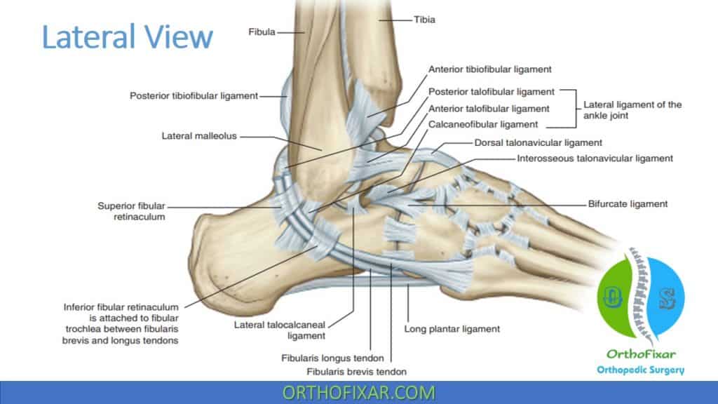
Lateral ankle ligaments Anatomy:
Lateral ankle ligaments function as a restraint to varus / inversion forces at ankle, besides maintaining lateral ankle stability, the lateral ankle ligaments play a significant role in maintaining rotational ankle stability.
- Anterior talofibular ligament (ATFL):
- A thickening of the anterior capsule extends from the anterior surface of the fibular malleolus, just lateral to the articular cartilage of the lateral malleolus, to just anterior to the lateral facet of the talus and to the lateral surface of the talar neck.
- It is an intracapsular structure and is approximately 2–5-mm thick and 10–12-mm long.
- The ATFL functions to resist ankle inversion in plantarflexion. Regardless of ankle position, the ATFL is usually the first ankle ligament to be torn in an inversion injury.
- Physical exam: anterior drawer test of the ankle in 20° of plantar flexion.
- Its Injury causes low ankle sprains.
- Calcaneofibular ligament (CFL):
- It is an extra-articular structure covered by the fibular (peroneal) tendons, is larger and stronger than the ATFL.
- It fans out at 10–40 degrees from the tip of the fibular malleolus to the lateral side of the calcaneus, paralleling the horizontal axis of the subtalar joint. This ligament effectively spans the ankle and subtalar joints, which have markedly different axes of rotation.
- Physical exam: inversion (supination) test and talar tilt test.
- Its injury is rare (low ankle sprain).
- Posterior talofibular ligament (PTFL):
- The ligament is coalescent with the joint capsule, and its orientation is relatively horizontal. Its attachment on the talus involves nearly the entire nonarticular portion of the posterior talus to the groove for the flexor hallucis longus (FHL) tendon, and anteriorly to the digital fossa of the fibula, which transmits the vessels that supply the talus and the fibula.
- It is the strongest of the lateral ligament complex, and serves to indirectly aid talofibular stability during dorsiflexion due to its anatomic location, where it can act as a true collateral ligament and prevent talar tilt into inversion.
- The PTFL is rarely injured except in severe ankle sprains.
- Physical exam: posterior drawer test of the ankle.
- Lateral Talocalcaneal Ligament (LTCL):
- It’s a short narrow ligamentous band that connects the lateral process of the talus to the lateral surface of the calcaneus.
- Located anterior and medial to calcaneofibular ligament.
- Its function is to stabilize the talocalcaneal joint.
- Anterior talofibular ligament (ATFL) is the weakest ankle ligament.
- Posterior talofibular ligament (PTFL) is the strongest ankle ligament.
In dorsiflexion, the PTFL is maximally stressed, and the CFL is taut, whereas the ATFL is loose. Conversely, in plantarflexion, the ATFL is taut, and the CFL and PTFL become loose.

Reference
- Frost HM, Hanson CA. Technique for testing the drawer sign in the ankle. Clin Orthop Relat Res. 1977 Mar-Apr;(123):49-51. PMID: 852190.
- Larkins LW, Baker RT, Baker JG. Physical Examination of the Ankle: A Review of the Original Orthopedic Special Test Description and Scientific Validity of Common Tests for Ankle Examination. Arch Rehabil Res Clin Transl. 2020 Jul 8;2(3):100072. doi: 10.1016/j.arrct.2020.100072. PMID: 33543095; PMCID: PMC7853358.
- Millers Review of Orthopaedics -7E Book
- Clinical Tests for the Musculoskeletal System 3rd Ed. Book.
- Lifetime product updates
- Install on one device
- Lifetime product support
- Lifetime product updates
- Install on one device
- Lifetime product support
- Lifetime product updates
- Install on one device
- Lifetime product support
- Lifetime product updates
- Install on one device
- Lifetime product support