Patellar Grind Test (Clarke Sign)
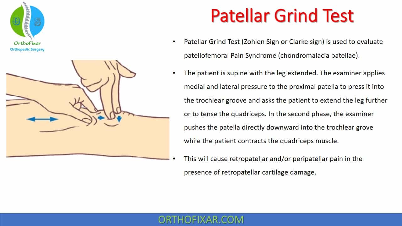
Patellar Grind Test (Clarke test or Zohlen Sign) is used to evaluate patellofemoral Pain Syndrome (chondromalacia patellae).
How do you perform the Patellar Grind Test?
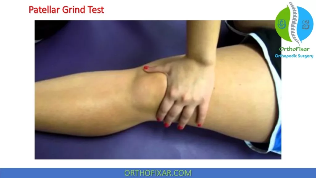
The Patellar Grind Test (Clarke test) is performed with the patient in supine position with the leg extended.
The examiner applies medial and lateral pressure to the proximal patella to press it into the trochlear groove and asks the patient to extend the leg further or to tense the quadriceps.
In the second phase, the examiner pushes the patella directly downward into the trochlear grove while asking the patient to contract the quadriceps muscle.
See Also: Patellar Glide Test
What does a positive Patellar Grind Test mean?
The quadriceps exerts a proximal pull on the patella, pressing it tightly against the trochlear groove. This will cause retropatellar and/or peripatellar pain in the presence of retropatellar cartilage damage.
Sensitivity & Specificity
A Validation study by Scott T Doberstein1 to evaluate the diagnostic value of the Clarke test in assessing Chondromalacia Patella, he found the sensitivity & specificity was:
- Sensitivity: 39 %
- Specificity: 67 %
This study concluded that the diagnostic validity values for the use of the Clarke test in assessing chondromalacia patella were unsatisfactory, supporting suggestions that it has poor diagnostic value as a clinical examination technique. Additionally, an extensive search of the available literature for the Clarke sign reveals multiple problems with the test, causing significant confusion for clinicians. Therefore, the use of the Clarke sign as a routine part of a knee examination is not beneficial, and its use should be discontinued.
Notes
As Clarke test is often positive in normal patients, one should repeat the procedure several times, increasing the pressure on the patella each time and comparing the results with those of the unaffected side.
To better localize the retropatellar damage, the knee should be tested in 30°, 60°, and 90° of flexion as well as in full extension.
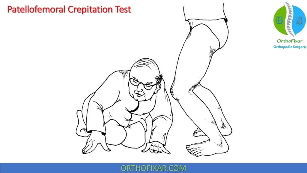
Patellofemoral Crepitation Test
Patellofemoral Crepitation Test is another test used in evaluation of chondromalacia (especially grades II and III)
The examiner kneels in front of the patient and asks the patient to crouch down or do a deep knee bend. The examiner listens for sounds posterior to the patella.
Crepitation (“snow ball crunch” sound) suggests severe chondromalacia (grades II and III). Painless cracking sounds like those that occur in almost everyone during the first or second deep knee bend have no significance. For this reason, the patient is asked to do several deep knee bends. Usually the insignificant cracking sounds will decrease in intensity.
In the absence of any audible retropatellar crepitation, the examiner may safely conclude that no severe retropatellar cartilage damage is present. However, the test results should not be used as a basis for far-reaching therapeutic decisions. They only provide information about the condition of the retropatellar cartilage. The crepitation test w ill be positive in m any patients with norm al knees.
Chondromalacia patella
The term chondromalacia patella has long been a catch-all category of anterior and, specifically, retropatellar knee pain.
The term chondromalacia was first used by Aleman in 1928. He used the term “chondromalacia posttraumatica patellae” to describe lesions of the patella found at surgery and thought to be caused by prior trauma.
With time, chondromalacia became an all-inclusive term with no agreed-on classification system or definitions of terms.
True chondromalacia refers to a softening of the cartilage on the posterior aspect of the patella and is estimated to occur in fewer than 20% of persons who present with anterior knee pain.
The syndrome is most common in the 12–35-year-old age group, and most studies show a predominance in females.
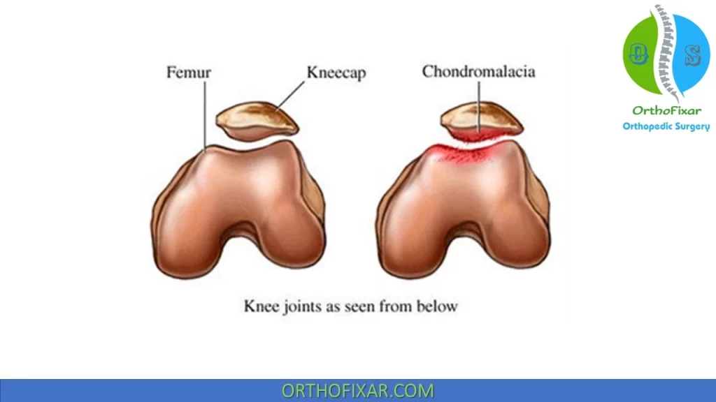
Two types of chondromalacia have been described:
- One type involves surface degeneration of the patella that is age dependent and most often asymptomatic.
- The other type involves basal degeneration. This type results from trauma and abnormal tracking of the patella and is symptomatic.
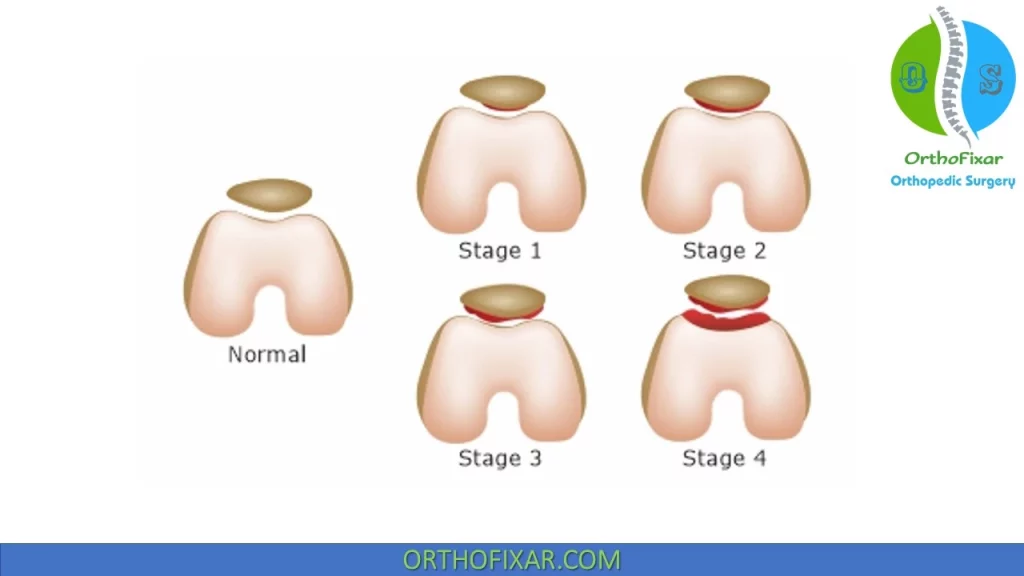
Chondromalacia patella Classification:
Chondromalacia is classified into four grades, according to the degree of degeneration noted arthroscopically:
- Grade 1: Closed disease. This grade is characterized by an intact joint surface that is spongy. The softening is
reversible. A blister or raised portion of the articular surface is seen. - Grade 2: Open disease. This grade is characterized by fissures that may or may not be obvious initially.
- Grade 3: Severe exuberant fibrillation or crabmeat appearance.
- Grade 4: The fibrillation is full-thickness and the erosive changes extend down to bone, which may be exposed. This is, in effect, OA and its extent depends on the size of the lesion.
References
- Doberstein ST, Romeyn RL, Reineke DM. The diagnostic value of the Clarke sign in assessing chondromalacia patella. J Athl Train. 2008 Apr-Jun;43(2):190-6. doi: 10.4085/1062-6050-43.2.190. PMID: 18345345; PMCID: PMC2267328.
- Ficat RP, Philippe J, Hungerford DS. Chondromalacia patellae: a system of classification. Clin Orthop Relat Res. 1979 Oct;(144):55-62. PMID: 535251.
- Aleman O: Chondromalacia post-traumatica patellae. Acta Chir Scand 63:149–190, 1928
- Holmes SW Jr, Clancy WG Jr. Clinical classification of patellofemoral pain and dysfunction. J Orthop Sports PhysioTherapy. 1998 Nov;28(5):299-306. doi: 10.2519/jospt.1998.28.5.299. PMID: 9809278.
- OUTERBRIDGE RE. The etiology of chondromalacia patellae. J Bone Joint Surg Br. 1961 Nov;43-B:752-7. doi: 10.1302/0301-620X.43B4.752. PMID: 14038135.
- Patellar Grind Test – Physiopedia
- Dutton’s Orthopaedic Examination, Evaluation, And Intervention 3rd Edition.
- Campbel’s Operative Orthopaedics 12th edition Book.
- Millers Review of Orthopaedics -7th Edition Book.
- Lifetime product updates
- Install on one device
- Lifetime product support
- Lifetime product updates
- Install on one device
- Lifetime product support
- Lifetime product updates
- Install on one device
- Lifetime product support
- Lifetime product updates
- Install on one device
- Lifetime product support