Special Test
Log Roll Test for Hip Pain Diagnosis

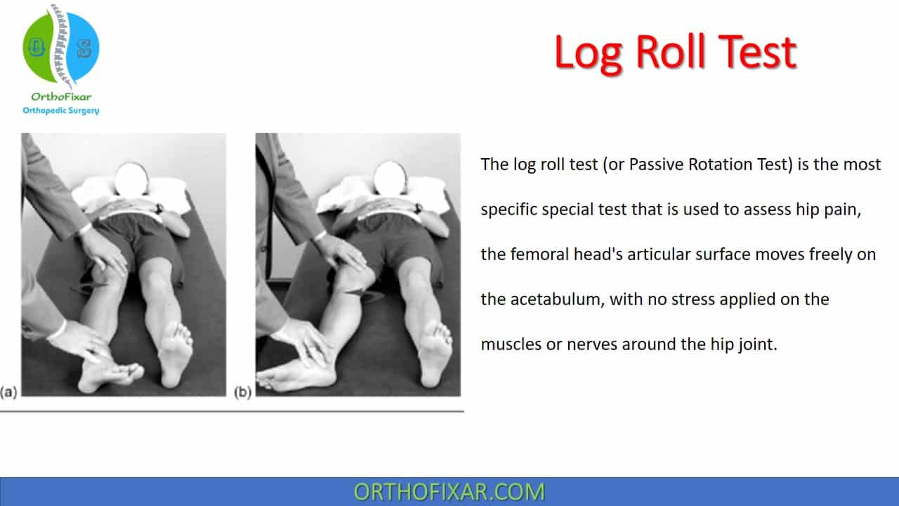
The log roll test (or Passive Rotation Test) is the most specific special test that is used to assess hip pain.
Where the femoral head’s articular surface moves freely on the acetabulum, with no stress applied on the muscles or nerves around the hip joint.
How do you do the log roll test?
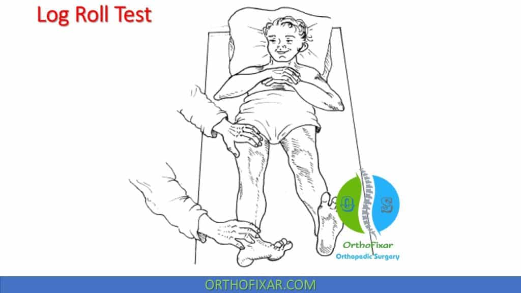
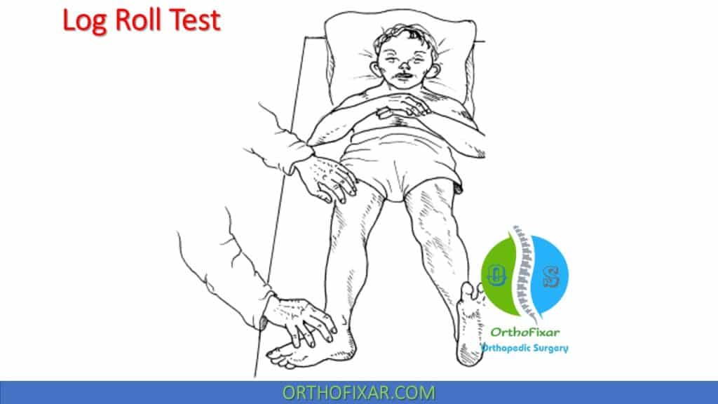
- With the patient supine, the examiner places one hand on the thigh and the other on the ankle and gently rolls the leg back and forth, thus alternately internally and externally rotating the hip.
- During this process, the joint surfaces of the femoral head move within the acetabulum without activating neural or myotendinous structures.
- The log roll test should be compared to the opposite hip.
What does a positive Log Roll Test mean?
Log Roll Test is positive when there is:
- Pain: refers to an intra-articular pathology.
- Clicking: refers to acetabular labral tear.
- Increased range of motion: refers to ligamentous laxity.


Sensitivity & Specificity
A systematic review of the diagnostic physical examination tests of the hip joint found that the log roll test hip fracture reliability (for femoral neck fractures) was:
- Sensitivity: 100%
- Specificity: 33%
Notes
- The log roll test is used to differentiate the pathologies inside the hip joint from those outside it.
- A negative log roll test does not exclude the hip as a source of symptoms, but a positive test give a high suspicious of hip pathology.
Related Anatomy
The hip articulation is a ball-and-socket joint formed between the head of the femur and the acetabulum of the pelvic bone.
- Structurally, the hip is suited for stability first, then mobility.
- The primary function of the hip is to support the weight of the head, arms, and trunk during the static erect posture and during dynamic activities such as ambulation, running, and stair climbing.
- In addition, the hip joint provides a pathway for the transmission of forces between the pelvis and the lower extremities.
- The hip joint is a marvel of physics, transmitting truly impressive loads, both tensile and compressive. For example,
during walking, the hip supports 1.3–5.8 times the body weight, and 4.5–8 times the body weight while running. - Finally, the hip joint functions to provide a wide range of lower limb movement.
- The hip joint is well designed to provide such an important service, provided that it is permitted to grow and develop normally.
The Femoral Head:
- The femoral head is composed of a trabecular bone core encased in a thin cortical bone shell.
- Both the trabecular framework of the head and its pliable hyaline cartilage covering contribute toward the shaping of the femoral head within the acetabulum during full weight-bearing.
- The femoral neck serves to extend the weight-bearing forces lateral and inferior to the joint fulcrum.
- The head of the femur is angled anteriorly, superiorly, and medially. The femoral neck is externally rotated with respect to the shaft.
- The angle that the femoral neck makes with the acetabulum is called the angle of anteversion/declination.
Acetabulum:
- The ilium, ischium, and pubis fuse together within the acetabulum and form a deep-seated depression in the lateral pelvis, which allows for the proximal transmission of weight from the axial skeleton to the lower extremity.
- The articular surface is covered by a thickened layer of hyaline cartilage, which thins near the center of the joint, and is absent over the acetabular notch, the area occupied by the ligamentum teres and obturator artery.
- The surface of the acetabulum faces laterally, inferiorly, and anteriorly.
- Around the periphery of the acetabulum is a thickened collar of fibrocartilage known as the acetabular rim, or labrum that further deepens the concavity and grasps the head of the femur.
- The diameter of the acetabulum is slightly less than that of the femoral head and results in an incongruous fit of the joint surfaces. This incongruity unloads the joint during partial weight-bearing (PWB), by allowing the femoral head to sublux laterally out of the cup, while in full weight-bearing, the femoral head is forced into the acetabulum.
See Also: Pelvic Anatomy

Reference
- J. W. Thomas Byrd, M.D. Evaluation of the Hip: History and Physical Examination. N Am J Sports Phys Ther. 2007 Nov; 2(4): 231–240. PMID: 21509142.
- Byrd JW. Evaluation of the hip: history and physical examination. N Am J Sports Phys Ther. 2007 Nov;2(4):231-40. PMID: 21509142; PMCID: PMC2953301.
- Rahman LA, Adie S, Naylor JM, Mittal R, So S, Harris IA. A systematic review of the diagnostic performance of orthopedic physical examination tests of the hip. BMC Musculoskelet Disord. 2013 Aug 30;14:257. doi: 10.1186/1471-2474-14-257. PMID: 23987589; PMCID: PMC3766647.
- Clinical Tests for the Musculoskeletal System 3rd Edition.
- Dutton’s Orthopaedic Examination, Evaluation, And Intervention 3rd Edition.
Angle Meter App for Android & iOS
- Lifetime product updates
- Install on one device
- Lifetime product support
Orthopedic FRCS VIVAs Quiz
- Lifetime product updates
- Install on one device
- Lifetime product support
Top 12 Best Free Orthopedic Apps
- Lifetime product updates
- Install on one device
- Lifetime product support
All-in-one Orthopedic App
- Lifetime product updates
- Install on one device
- Lifetime product support