Hoffa’s Syndrome
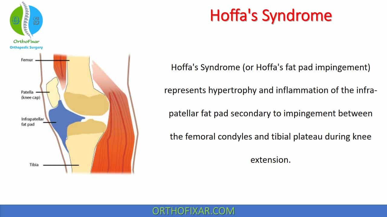
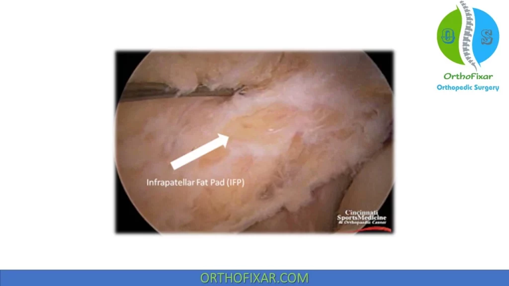
Hoffa’s Syndrome (or Hoffa’s fat pad impingement) represents hypertrophy and inflammation of the infra-patellar fat pad secondary to impingement between the femoral condyles and tibial plateau during knee extension.
It’s sometimes called Infrapatellar fat pad syndrome. This syndrome was first described by Hoffa in 1904.
See Also: Homans Sign
Fat Pads Anatomy
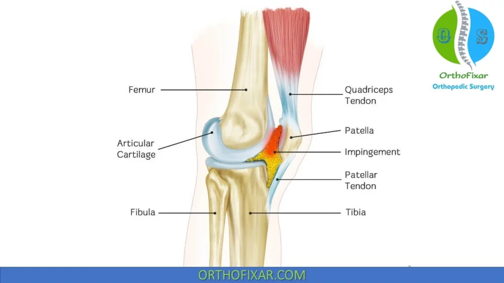
There are three fat pads located at the anterior knee:
- The quadriceps fat pad,
- The prefemoral fat pad,
- The infrapatellar fat pad (Hoffa’s fat pad).
Infrapatellar fat pad, which is called Hoffa’s fat pad, is located between the patella ligament (anteriorly) and the anterior joint capsule (posteriorly).
The functions of the fat pads include the following:
- Synovial fluid secretion,
- Joint stability,
- Neurovascular supply,
- Occupiers of dead space.
Hoffa’s Syndrome Causes
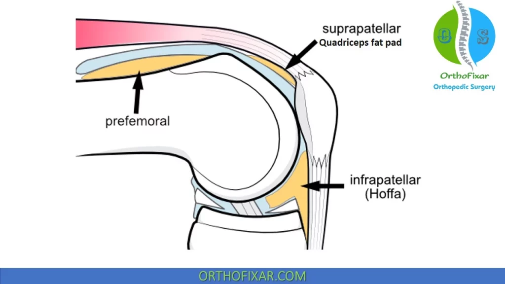
- The main cause is the impingement between the femoral condyles and tibial plateau during knee extension.
- Direct trauma and overuse have also been attributed as causes.
- Irritation also can be produced by a posterior tilt of the inferior pole of patella.
Hoffa’s fat pad impingement Symptoms
Symptoms include anterior knee pain that is inferior to the pole of the patella. Pain is exacerbated by knee extension, particularly hyperextension, but not by knee flexion.
Inspection may reveal inferior patellar edema and associated tenderness of the fat pad when palpated through the tendon.
In chronic cases, recurrent hydrarthrosis may present, together with joint weakening and subpatellar discomfort.
Direct palpation of the fat pad on either side of the patellar tendon as the knee is brought from flexion into full extension is painful if the fat pad is inflamed.
A diagnostic Hoffa’s Syndrome test, termed the bounce test (eliciting pain with passive knee hyperextension), is sometimes useful.
One must be careful to look for other causes of inflammation, such as knee OA, before concluding that the fat pad impingement is the primary source of pain.
Radiology
Plain radiographs are invariably negative.
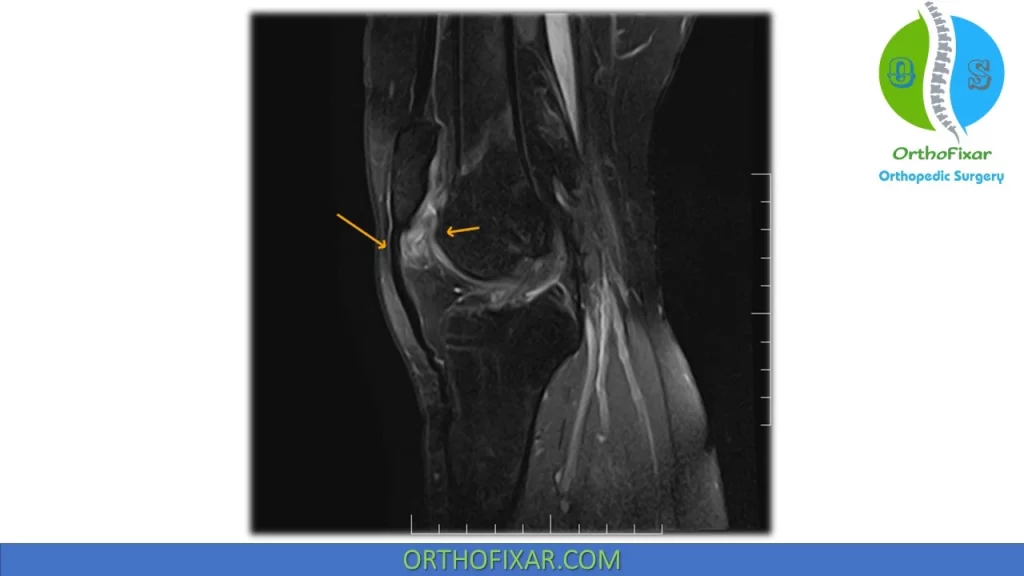
MRI has high sensitivity in Hoffa’s fat pad, abnormalities findings of the fat pad can be noted on MRI:
- T1-weighted sequences showed the fibrotic trabeculae of the pad.
- T2-weighted sequences demonstrated liquid infiltration in the pad and various synovial recesses.
Hoffa’s fat pad Syndrome Treatment
Hoffa’s syndrome treatment includes:
- Rest,
- Ice,
- Anti-inflammatory medications NSAIDs,
- Iontophoresis or phonophoresis.
- Local corticosteroid injections into the fat pad are preferred by some physicians because they can be both diagnostic and therapeutic.
Biomechanical interventions include addressing the causes of hyperextension through orthotic interventions, such as heel lifts or taping the superior pole posteriorly and holding the patella in a superior glide with tape.
In recalcitrant cases, surgical resection of portions of the fat pad is indicated (arthroscopic resection).
Article Summary
Hoffa’s Syndrome is a condition that causes inflammation and enlargement of the infrapatellar fat pad, located below the kneecap. It is caused by impingement between the femoral condyles and tibial plateau during knee extension, as well as trauma and overuse. Symptoms include anterior knee pain that worsens with knee extension, inferior patellar edema, and tenderness. Diagnosis is typically done through MRI. Treatment options include rest, ice, anti-inflammatory medication, corticosteroid injections, and surgical resection in severe cases. Biomechanical interventions such as orthotic interventions and taping can also be helpful.
References
- Hoffa A: The influence of the adipose tissue with regard to the pathology of the knee joint. J Am Med Assoc 43:795–796, 1904.
- Jacobson JA, Lenchik L, Ruhoy MK, Schweitzer ME, Resnick D. MR imaging of the infrapatellar fat pad of Hoffa. Radiographics. 1997 May-Jun;17(3):675-91. doi: 10.1148/radiographics.17.3.9153705. PMID: 9153705.
- Draghi F, Ferrozzi G, Urciuoli L, Bortolotto C, Bianchi S. Hoffa’s fat pad abnormalities, knee pain and magnetic resonance imaging in daily practice. Insights Imaging. 2016 Jun;7(3):373-83. doi: 10.1007/s13244-016-0483-8. Epub 2016 Mar 21. PMID: 27000624; PMCID: PMC4877349.
- Morini G, Chiodi E, Centanni F, Gattazzo D. Malattie del corpo adiposo di Hoffa: Risonanza Magnetica e chirurgia a confronto [Hoffa’s disease of the adipose pad: magnetic resonance versus surgical findings]. Radiol Med. 1998 Apr;95(4):278-85. Italian. PMID: 9676203.
- Safran MR, Fu FH. Uncommon causes of knee pain in the athlete. Orthop Clin North Am. 1995 Jul;26(3):547-59. PMID: 7609965.
- Holmes SW Jr, Clancy WG Jr. Clinical classification of patellofemoral pain and dysfunction. J Orthop Sports Phys Ther. 1998 Nov;28(5):299-306. doi: 10.2519/jospt.1998.28.5.299. PMID: 9809278.
- Saxena S, Patel DD, Shah A, Doctor M. Fat Chance for Hidden Lesions: Pictorial Review of Hoffa’s Fat Pad Lesions. Indian J Radiol Imaging. 2021 Nov 30;31(4):961-974. doi: 10.1055/s-0041-1739383. PMID: 35136510; PMCID: PMC8817800.
- Dutton’s Orthopaedic Examination, Evaluation, And Intervention 3rd Edition.
- Lifetime product updates
- Install on one device
- Lifetime product support
- Lifetime product updates
- Install on one device
- Lifetime product support
- Lifetime product updates
- Install on one device
- Lifetime product support
- Lifetime product updates
- Install on one device
- Lifetime product support