Distal Femoral Osteotomy
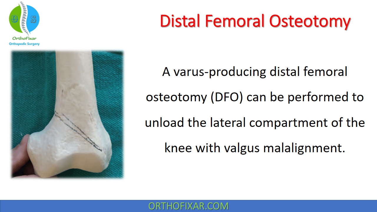
A varus-producing distal femoral osteotomy (DFO) can be performed to unload the lateral compartment of the knee with valgus malalignment.
The use of high tibial osteotomy and Distal Femoral Osteotomy for the treatment of gonarthrosis follows the theory of limb realignment in the coronal plane in order to unload the affected knee compartment and transfer the weightbearing forces through the healthy knee compartment.5,8 Valgus-producing osteotomies to unload the medial compartment are typically performed with an HTO (medial opening wedge or lateral closing wedge), whereas varus producing-osteotomies to unload the lateral compartment are performed with a distal femoral osteotomy(medial opening wedge or lateral closing wedge) or lateral opening wedge HTO.
This article describes the technique of a medial closing wedge distal femoral osteotomy for lateral gonarthrosis.
See Also: High Tibial Osteotomy
Distal Femoral Osteotomy Indications
- Gonarthrosis isolated to the lateral compartment and valgus malalignment
- Primary femoral deformity
- Large correction (ie, > 12 degrees of valgus deformity)
- Lateral opening wedge HTO can be considered for small corrections, with the advantage of correcting the deformity in extension and flexion; distal femoral osteotomy only corrects the deformity in extension.
Contraindications
- Contraindications for a distal femoral osteotomy include advanced age, stiffness, tricompartmental disease, and
inflammatory arthritis.
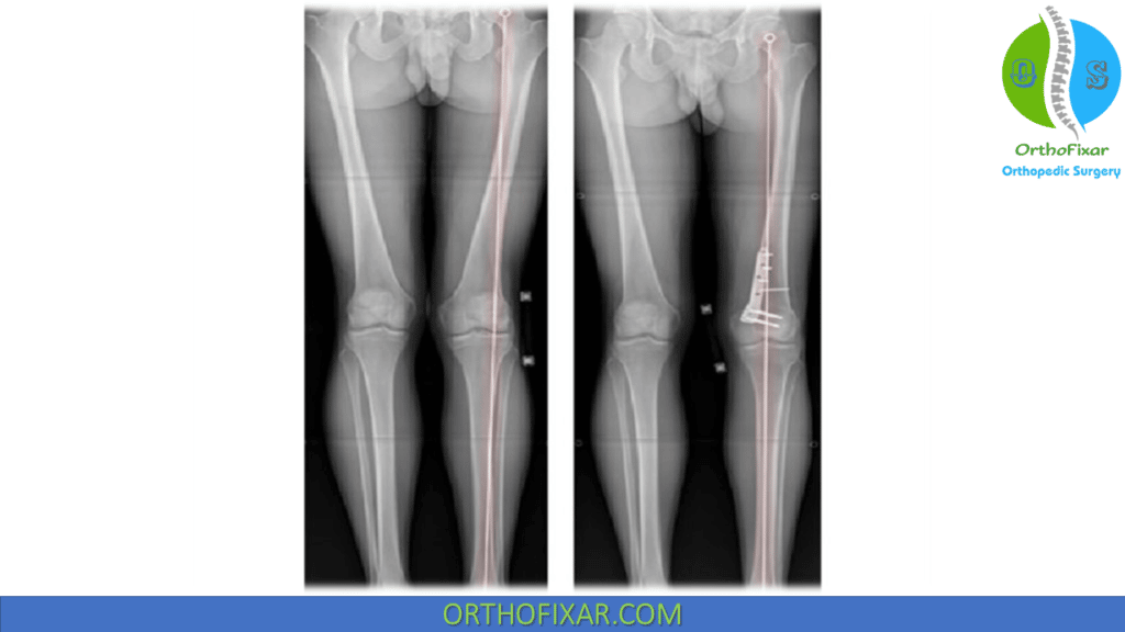
Medial Closing Wedge Distal Femoral Osteotomy
Patients with valgus alignment with the weightbearing axis extending through a diseased lateral compartment are candidates for a varus-producing procedure. For small corrections less than 10 mm, it’s preferred to correct below the knee with a lateral opening tibial osteotomy.
The indications for varus-producing osteotomies include isolated lateral compartment arthrosis with the medial and patellofemoral compartments having Outerbridge changes of grade II or less. The patient should be physiologically young, preferably with a healthy body mass index, and a nonsmoker, with more than 90 degrees knee flexion. Patients requiring larger corrections, including those with primarily femoral-sided valgus deformity will benefit from medial closing wedge distal femoral osteotomy.
On the femoral side, a lateral opening or medial closing procedure can achieve the desired correction; the advantages of medial closing are that:
- the hardware is less prominent medially,
- the closing wedge technique is stable,
- union occurs quickly.
The image intensifier is brought in from the affected side, and the surgeon works from the opposite (medial) side. A tourniquet is used as desired, and preoperative antibiotics are given routinely.
The incision is medial to the midline and longitudinal to facilitate any future reconstructions. The incision extends from the distal pole of the patella proximally for 15 cm. After dissection through the skin and subcutaneous tissue, the lower border of the vastus medialis is identified; in order to mobilize the vastus, the medial patellofemoral ligament is incised and later repaired. The vastus medialis is mobilized off the intermuscular septum, taking care to cauterize any perforating vessels. The distal two fifths of the medial femur can be safely exposed before encountering the adductor canal and the femoral artery.
See Also: Anteromedial Approach to Femur
A blunt retractor (Hohmann, Bennett, or curved knee) is placed beneath the vastus to retract it anteriorly. The intermuscular septum is carefully elevated off the PMC of the femur at the supracondylar level at the proposed osteotomy site, and a blunt Hohmann retractor is placed across the posterior femur directly on the bone to prevent neurovascular injury during the osteotomy. The periosteum is left intact to maintain vascularity. The hardware can then be trialed on the medial femur with fluoroscopic guidance.
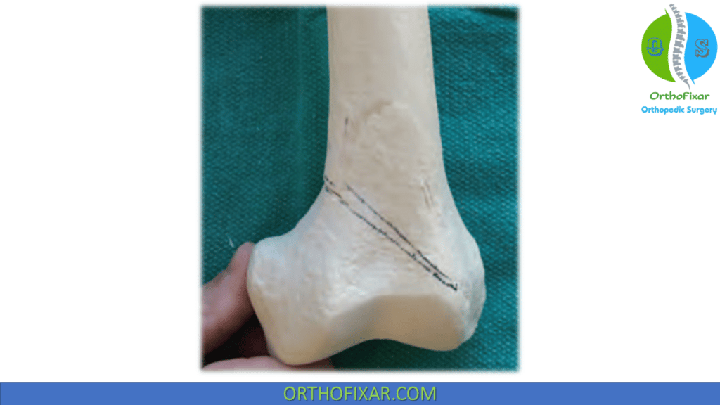
The plate is lined up with the anterior aspect of the plate flush with the anterior cortex of the femur. There is room for 4 screws above and below the osteotomy. The lower cut of the osteotomy should be approximately 10 mm above the superior extent of the trochlea; a small arthrotomy may be necessary to palpate this landmark. The proposed osteotomy is then marked using electrocautery on the medial cortex perpendicular to the long axis of the femur.
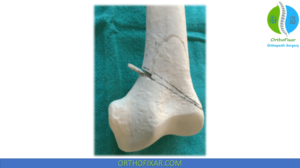
To make the osteotomy close with cortical contact, one should have both limbs of the osteotomy of equal length with the apex 10 mm medial to the lateral cortex. The apex should be below the level of the medial epicondyle because the MCL will provide additional restraint to translation of the osteotomy. K-wires can be used as cutting guides, being sure to account for the thickness of the saw blade when planning the wedge.
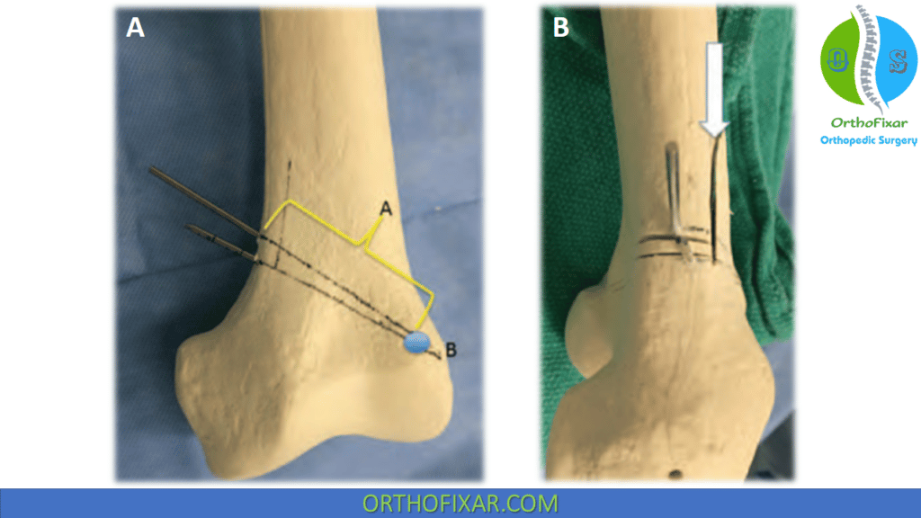
B: On the lateral view, an optional biplane cut is demonstrated (arrow)
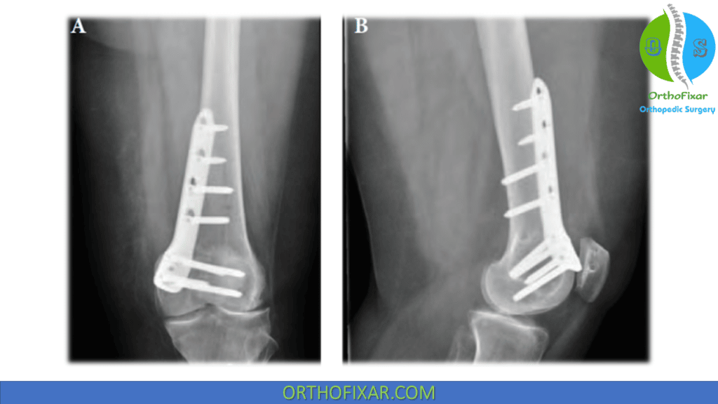
While using the saw, use plenty of irrigation to avoid heat build-up on the blade. The posterior Hohmann retractor should be right in line with the saw at all times. A series of osteotomes can be used to complete the cuts as needed. The bone wedge is then removed. A biplanar cut can be performed as an option to increase the stability and surface area of the osteotomy. Fixation is then achieved using standard fracture fixation techniques to achieve compression across the osteotomy site.
References
- Puddu G, Cipolla M, Cerullo G, Franco V, Giannì E. Osteotomies: the surgical treatment of the valgus knee. Sports Med Arthrosc Rev. 2007 Mar;15(1):15-22. doi: 10.1097/JSA.0b013e3180305c76. PMID: 17301698.
- Healy WL, Anglen JO, Wasilewski SA, Krackow KA. Distal femoral varus osteotomy. J Bone Joint Surg Am. 1988;70(1):102-109.
- Stahelin T, Hardegger F, Ward JC. Supracondylar osteotomy of the femur with use of compression. Osteosynthesis with a malleable implant. J Bone Joint Surg Am. 2000;82(5):712-722.
- Wang JW, Hsu CC. Distal femoral varus osteotomy for osteoarthritis of the knee. Surgical technique. J Bone Joint Surg Am. 2006;88(suppl 1 pt 1):100-108.
- Successful Return to Sport Following Distal Femoral Varus Osteotomy – Scientific Figure on ResearchGate. Available from: https://www.researchgate.net/figure/A-Standing-long-leg-radiograph-displaying-valgus-deformity-of-the-left-knee-with-a_fig3_322008207 [accessed 25 Sep, 2022]
- Lifetime product updates
- Install on one device
- Lifetime product support
- Lifetime product updates
- Install on one device
- Lifetime product support
- Lifetime product updates
- Install on one device
- Lifetime product support
- Lifetime product updates
- Install on one device
- Lifetime product support