Infant Hip Ultrasound

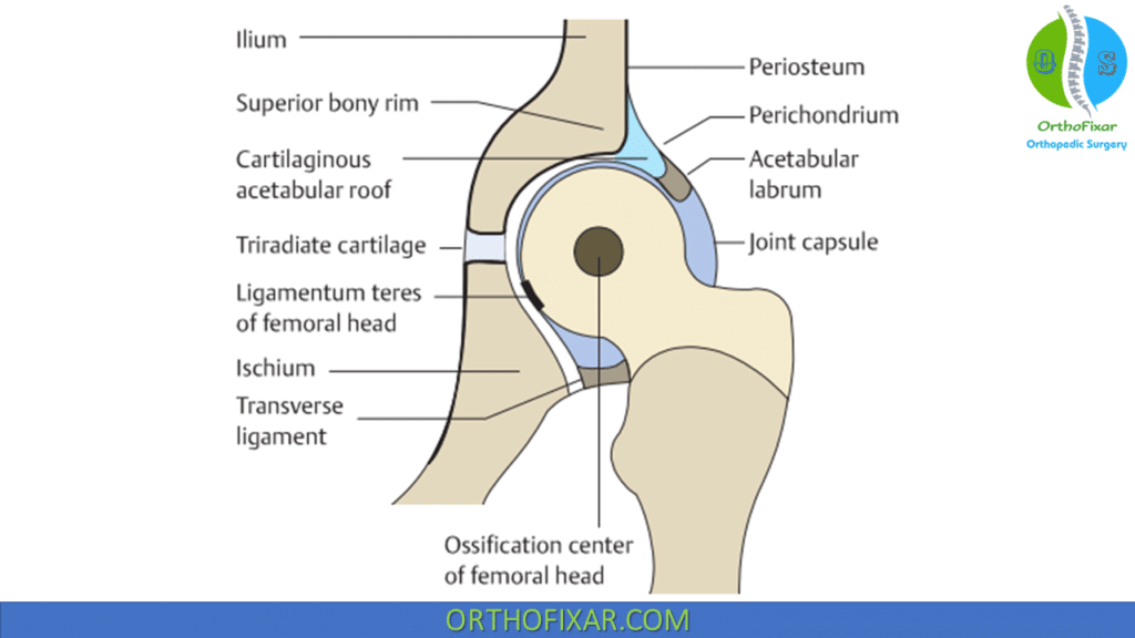
Infant Hip Ultrasound, a non-invasive, radiation-free imaging modality, has become the diagnostic gold standard for assessing hip joint integrity and alignment in infants. It allows dynamic assessment and visualization of cartilaginous and non-ossified hip structures, which are not discernible with conventional radiography in the first few months of life.
The diagnostic accuracy of infant hip ultrasound is highly dependent on both technique and interpretation, requiring a clear understanding of the intricate hip anatomy and morphological changes associated with age and development.
Infant Hip US is the preferred modality for evaluating the hip in infants aged less than 6 months.

Infant Hip Ultrasound Scanning Technique
Graf Method
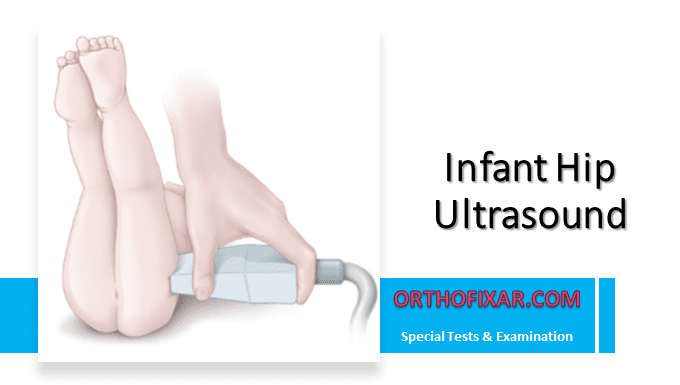
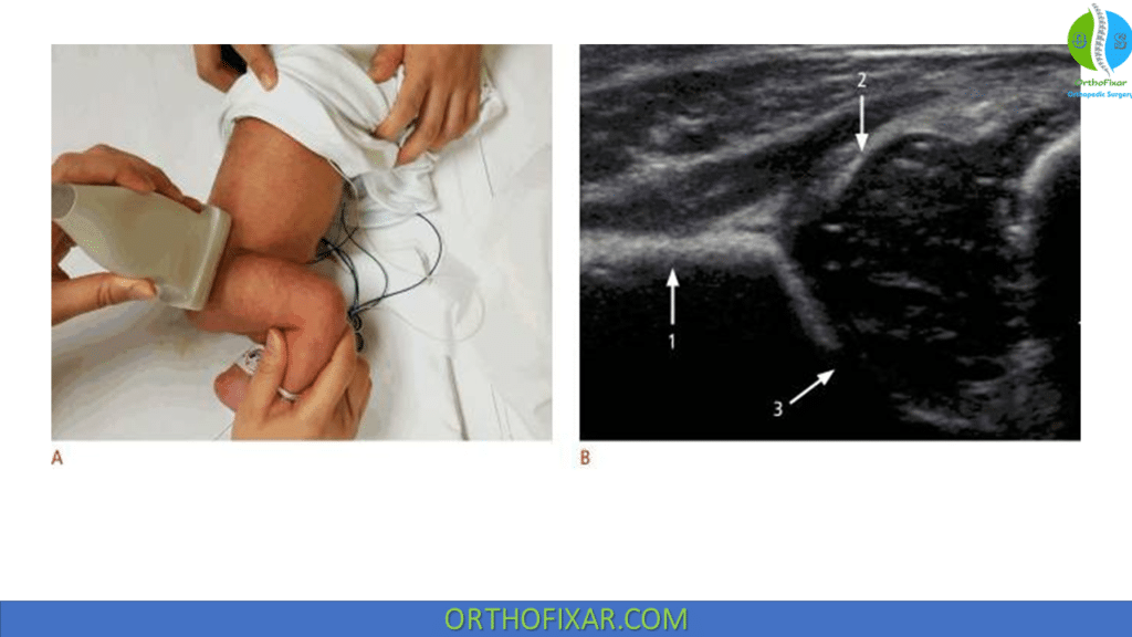
Graf method for infant hip ultrasound is a static morphologic evaluation of the infant hip. The infant is scanned in a lateral decubitus position with the examiner on the right side. The acetabulum appears on the right side of the image, the greater trochanter on the left side.
The hip joint is evaluated in a standard coronal plane with a linear array probe. Three key landmarks should be visualized in this plane:
- The inferior border of the ilium in the acetabular fossa.
- The central weight-bearing zone of the acetabular roof.
- The acetabular labrum.
The coronal view of the hip can be captured with the infant’s hip in either a standard neutral position, typically between 15° and 20° flexion, or a more flexed position. The ultrasound transducer is positioned in alignment with the coronal plane of the body. It is then moved back and forth from this foundational position to delineate the rounded configuration of the hip joint.

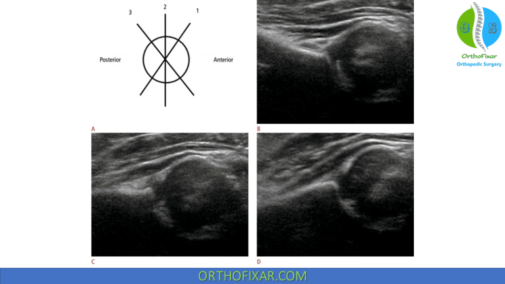
The image can be manipulated by slightly rotating the superior edge of the transducer posteriorly by an angle between 10° to 15° into an oblique coronal plane. This action gives the ilium a straight appearance. A standard plane of the hip is indicated if the sonogram illustrates a straight contour of the iliac wing, visibility of the triradiate cartilage, and a conspicuous acetabular labrum.
However, in cases of hip dislocation, the femoral head’s lateral and posterior displacement obstructs the view of both the femoral head and the acetabulum’s center in the standard plane. Hence, when the displaced femoral head is traced, the ultrasound plane deviates from the standard plane. The visible sectional plane is often posterior due to the femoral head’s direction of displacement.

The American College of Radiology suggests performing a standard ultrasound examination in the transverse view, with the hip flexed at a 90° angle. In this perspective, the femoral shaft can be seen anteriorly, ending at the femoral head, which is resting on the ischium.
To detect a potentially dislocatable hip, the transducer is positioned in a posterolateral position while performing the Ortolani and Barlow maneuvers. If the connection between the posterior acetabulum and the femoral head shifts under gentle stress, it indicates instability in the hip.

Alpha (α) and Beta (β) Angles:
All angles are measured in relation to the “baseline,” which is tangent to the lateral border of the ilium. Two more reference lines are drawn along the bony roof of the acetabulum (acetabular roof line) and along the acetabular labrum (labral line}:
- The acetabular roof line is drawn from the superior bony rim of the acetabulum to the lower edge of the ilium in the acetabular fossa.
- The labral line runs from the labrum to the superior bony rim of the acetabulum or to the transition point from the convexity of the superior bony rim to the concave acetabular roof.
The intersections of the acetabular roof line and the labral line form the alpha (α) and beta (β) angles of the hip:
- The α angle is formed by the baseline and acetabular roof line and reflects the bony coverage of the femoral head.
- The β angle, formed by the baseline and labral line, is a criterion for evaluating the cartilaginous acetabular roof.
See Also: Developmental Dysplasia of the Hip

Graf Classification System of DDH
Graf classified developmental dysplasia of the hip on the Basis of the sonographic angles of the hip:
| Class | Alpha Angle | Beta Angle | Description | Treatment |
|---|---|---|---|---|
| I | > 60° | < 55° | Normal | None |
| II | 43-60° | 55-77° | Delayed Ossification | Variable |
| III | < 43° | > 77° | Lateralization | Palvik Harness |
| IV | Unmeasurable | Unmeasurable | Dislocated | Pavlik harness/closed vs. open reduction |
See Also: Developmental Dysplasia of the Hip Risk Factors
References
- Kang YR, Koo J. Ultrasonography of the pediatric hip and spine. Ultrasonography. 2017 Jul;36(3):239-251. doi: 10.14366/usg.16051. Epub 2017 Feb 22. PMID: 28372341; PMCID: PMC5494873.
- Graf R. Sonographie der Sauglingshufte. 5th ed. Stuttgart: Thieme; 2000
- Graf R. Hip Sonograpby. Diagnosis and Management of Infant Hip Dysplasia. 2nd eel Berlin Heidelberg: Springer; 2006
- Tachdjian Pediatric Orthopaedics 5th book.
- Lifetime product updates
- Install on one device
- Lifetime product support
- Lifetime product updates
- Install on one device
- Lifetime product support
- Lifetime product updates
- Install on one device
- Lifetime product support
- Lifetime product updates
- Install on one device
- Lifetime product support