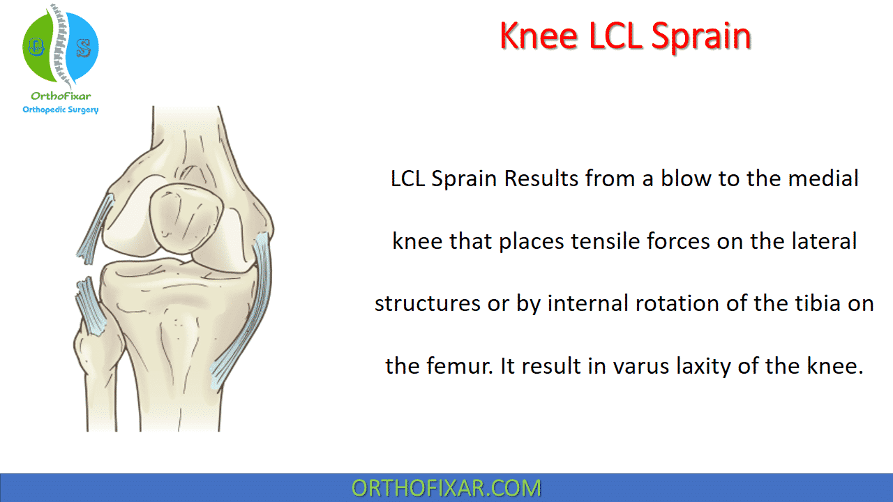
A Knee LCL Sprain is an injury to the lateral collateral ligament (LCL), one of the four major stabilizing ligaments of the knee. The LCL extends from the lateral femoral epicondyle to the fibular head and primarily resists varus stress (forces pushing the knee outward). It also contributes to controlling external tibial rotation and stabilizing the posterolateral aspect of the knee.
Unlike the medial collateral ligament (MCL), isolated LCL injuries are relatively uncommon because the ligament is protected by surrounding structures. However, LCL injuries frequently occur in association with posterolateral corner (PLC) injuries, cruciate ligament tears, or knee dislocations.
Prompt diagnosis is essential because untreated LCL injuries, particularly high-grade tears, may lead to chronic knee instability, pain, and early degenerative arthritis.
Related Anatomy
The lateral collateral ligament (LCL) does not attach to the joint capsule or meniscus. It’s a cordlike structure arises from the lateral femoral epicondyle, sharing a common site of origin with the lateral joint capsule, and inserts on the proximal aspect of the fibular head.
The LCL is the primary restraint against varus forces when the knee is in the range between full extension and 30 degrees of flexion. This structure also provides a primary restraint against external tibial rotation and a secondary restraint against internal rotation of the tibia on the femur.
The blood supply is from superolateral and inferolateral geniculate arteries.
See Also: Knee Ligaments Anatomy

Mechanism of Injury
Most LCL sprains occur following a force applied to the medial side of the knee, producing excessive varus stress.
Common mechanisms include:
- Contact sports (football, rugby, soccer)
- Skiing injuries
- Motor vehicle accidents
- Twisting injuries
- Hyperextension trauma
- Knee dislocation
High-energy trauma often causes combined injuries involving:
- ACL
- PCL
- Posterolateral corner
- Menisci
Risk Factors
Risk factors include:
- Contact sports participation
- Previous knee ligament injuries
- Poor neuromuscular control
- Inadequate rehabilitation
- High-speed pivoting activities
- Knee hyperextension injuries
LCL Sprain Classification
LCL Sprain is classified based on the lateral joint opening as compared with the normal contralateral knee
with varus stress:
- Grade I: 0-5 mm lateral opening.
- Grade II: 6-10 mm lateral opening.
- Grade III: >10 mm lateral opening without an endpoint.
It’s also classified based on the amount of ligamentous disruption:
- Grade I: minimal tear.
- Grade II: partial tear.
- Grade III: complete tear.
See Also: MCL Sprain
LCL Sprain Symptoms & Signs
Clinical presentation depends on injury severity.
Common symptoms include:
- Lateral knee pain
- Tenderness along the ligament
- Swelling
- Bruising
- Difficulty walking
- Knee instability
- Feeling that the knee “gives way”
- Pain during cutting or pivoting movements
- Reduced range of motion
Patients with associated PLC injuries often complain of rotational instability during walking.
Physical Examination
A complete ligamentous examination is essential.
Inspection
Look for:
- Swelling
- Ecchymosis
- Varus knee alignment
- Gait abnormalities
Palpation
Tenderness is usually localized between:
- Lateral femoral epicondyle
- Fibular head
Range of Motion
Assess:
- Active ROM
- Passive ROM
- Pain with knee flexion and extension
Special Tests
Varus Stress Test
The most important examination.
Performed at:
- 30° knee flexion
- Isolates the LCL
- Full extension
- Evaluates combined ligament injuries
Positive findings:
- Lateral joint opening
- Pain
- Soft endpoint
Dial Test
Useful for identifying:
- Posterolateral corner injuries
- Combined LCL injuries
Increased external tibial rotation compared with the opposite knee suggests PLC injury.
External Rotation Recurvatum Test
Assesses:
- PLC injuries
- Combined ligament injuries
Posterolateral Drawer Test
Evaluates posterolateral instability.
See Also: Varus Stress Test
Imaging
Plain Radiographs
Recommended views include:
- AP
- Lateral
- Tunnel
- Merchant view
Radiographs help identify:
- Fractures
- Fibular head avulsion (Arcuate sign)
- Varus alignment
- Joint space widening
Stress radiographs may quantify lateral compartment opening.
MRI
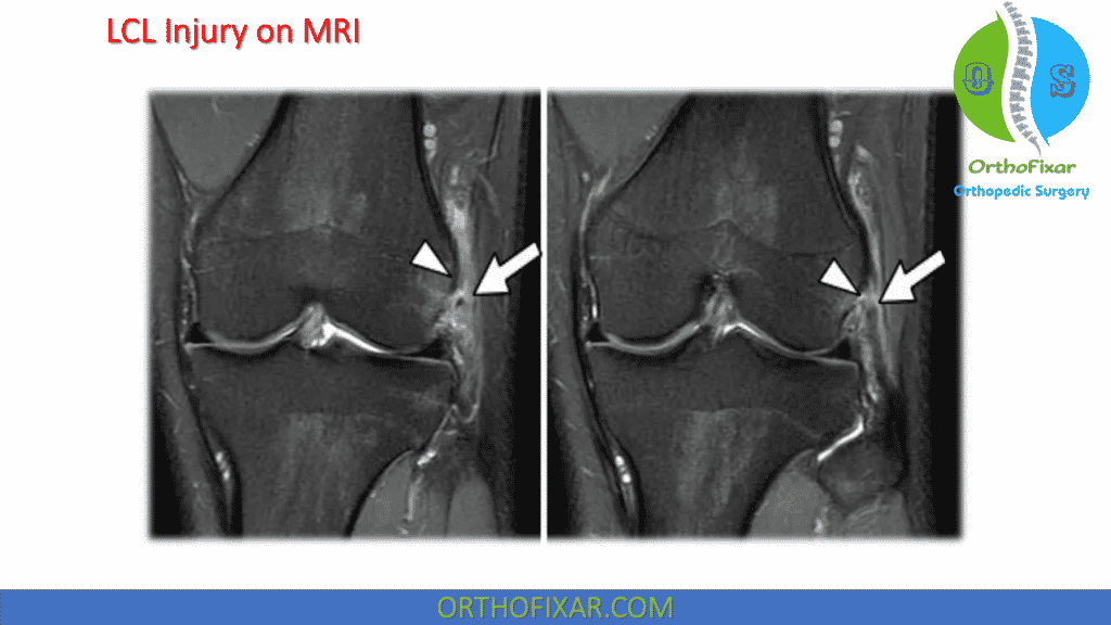
MRI is the gold standard for evaluating LCL injuries.
MRI identifies:
- Partial tears
- Complete ruptures
- Associated ACL/PCL injuries
- Meniscal tears
- Bone bruises
- PLC injuries
- Cartilage damage
MRI has excellent sensitivity and specificity for diagnosing multiligament knee injuries.
Ultrasound
Dynamic musculoskeletal ultrasound may detect:
- Partial tears
- Complete tears
- Ligament thickening
- Hematoma
However, it is operator-dependent and less comprehensive than MRI.
Knee LCL Sprain Treatment
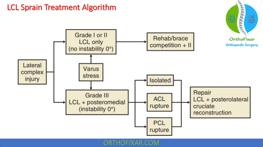
Treatment depends on injury grade and associated ligament damage.
Nonoperative Management
Usually indicated for:
- Grade I injuries
- Most Grade II injuries
- Stable isolated LCL injuries
Treatment includes:
Initial Management (PRICE)
- Protection
- Relative rest
- Ice
- Compression
- Elevation
NSAIDs may help reduce pain and inflammation during the acute phase, although prolonged use should be avoided if possible due to potential effects on tissue healing.
Knee Bracing
Functional hinged knee braces help:
- Reduce varus stress
- Improve confidence
- Protect healing tissue
Typically worn for:
- 2–6 weeks
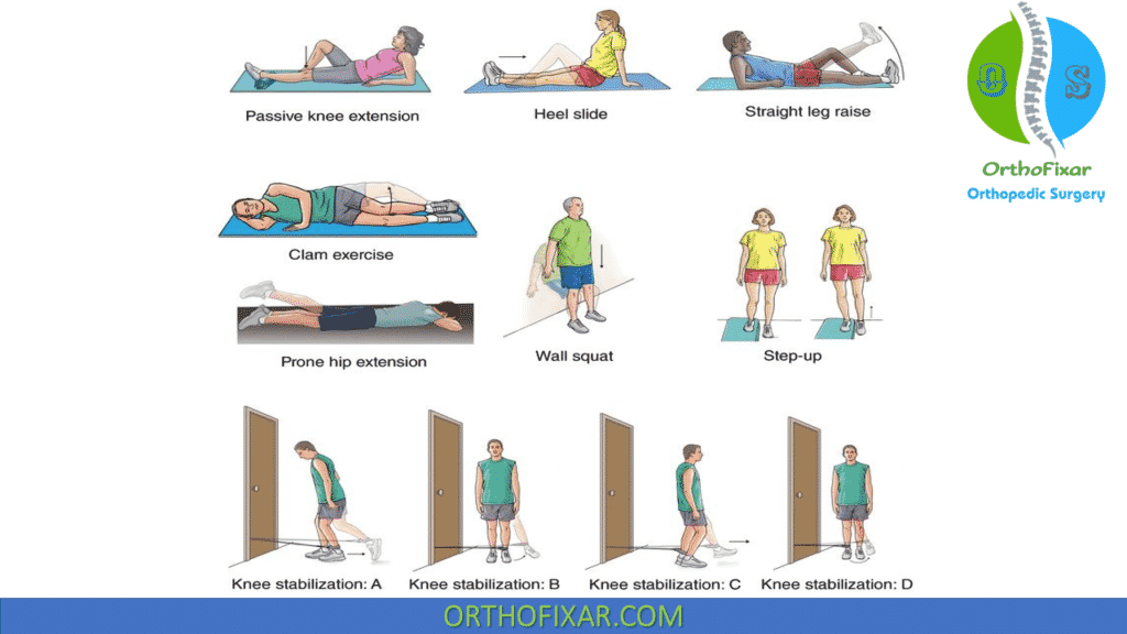
Physical Therapy
Rehabilitation focuses on:
- Early range of motion
- Quadriceps strengthening
- Hamstring strengthening
- Hip abductor strengthening
- Core stability
- Proprioception training
- Balance exercises
- Sport-specific drills
Surgical Management
Surgery is generally recommended for:
- Grade III tears
- Combined ligament injuries
- PLC injuries
- Knee dislocations
- Chronic instability
- Failed conservative treatment
Procedures include:
Primary Repair
Most appropriate for:
- Acute proximal or distal avulsion injuries
- Good tissue quality
LCL Reconstruction
Often preferred for:
- Chronic tears
- Mid-substance ruptures
- Poor tissue quality
Common graft options:
- Semitendinosus autograft
- Achilles tendon allograft
- Tibialis anterior allograft
Modern anatomic reconstruction techniques provide superior restoration of knee biomechanics compared with older non-anatomic procedures.
Rehabilitation Protocol
Phase 1 (Weeks 0–2)
Goals:
- Control swelling
- Reduce pain
- Restore extension
- Partial weight bearing if indicated
Exercises:
- Quadriceps sets
- Straight-leg raises
- Ankle pumps
- Patellar mobilization
Phase 2 (Weeks 2–6)
Goals:
- Improve ROM
- Normalize gait
- Increase strength
Exercises:
- Mini squats
- Stationary cycling
- Step-ups
- Closed-chain strengthening
Phase 3 (Weeks 6–12)
Goals:
- Restore strength
- Improve neuromuscular control
Exercises:
- Single-leg balance
- Lunges
- Resistance training
- Agility drills
Phase 4 (3–6 Months)
Goals:
- Return to sports
- Maximize functional stability
Activities:
- Running progression
- Plyometrics
- Cutting drills
- Sport-specific training
Recovery Time
Approximate recovery times:
| Injury Grade | Recovery |
|---|---|
| Grade I | 2–4 weeks |
| Grade II | 6–8 weeks |
| Grade III (nonoperative) | 8–12 weeks |
| After reconstruction | 6–9 months |
Return-to-play decisions should be based on:
- Full ROM
- Strength ≥90% of the contralateral limb
- Absence of instability
- Successful functional testing
Complications
Possible complications include:
- Chronic varus instability
- Persistent pain
- Knee stiffness
- Peroneal nerve injury
- Recurrent instability
- Failure of reconstruction
- Early osteoarthritis
- Missed posterolateral corner injury
Prognosis
The prognosis is generally excellent for isolated Grade I and Grade II injuries treated appropriately, with most patients returning to their previous activity level.
Grade III injuries have more variable outcomes and often require surgical reconstruction, particularly when associated with PLC or cruciate ligament injuries. Early recognition and appropriate management significantly improve long-term knee stability and functional outcomes.
Prevention
Strategies to reduce the risk of LCL injury include:
- Regular lower extremity strengthening
- Neuromuscular training programs
- Balance and proprioception exercises
- Proper landing mechanics
- Sport-specific conditioning
- Adequate warm-up before activity
- Appropriate protective equipment in contact sports
Frequently Asked Questions (FAQs)
Can an LCL sprain heal without surgery?
Yes. Most isolated Grade I and Grade II LCL sprains heal successfully with conservative treatment, including bracing, physical therapy, and gradual return to activity. Complete Grade III tears or injuries associated with other knee ligaments often require surgical management.
How long does an LCL sprain take to heal?
Recovery depends on injury severity. Mild sprains usually recover within 2–4 weeks, moderate injuries in 6–8 weeks, while complete tears treated surgically may require 6–9 months before returning to sports.
Can you walk with an LCL tear?
Many patients can still walk with a mild or partial LCL injury, although pain and instability may be present. Complete tears often produce significant instability and difficulty with weight-bearing, especially during turning or pivoting.
What is the difference between an LCL and MCL injury?
The LCL is located on the outside (lateral aspect) of the knee and resists varus forces, whereas the MCL is located on the inside (medial aspect) and resists valgus forces. MCL injuries are considerably more common than isolated LCL injuries.
Is MRI necessary for an LCL sprain?
MRI is recommended when a high-grade injury, combined ligament injury, or posterolateral corner injury is suspected. It provides detailed assessment of the LCL and associated structures and helps guide treatment decisions.
References
- Murakami Y, Ochi M, Ikuta Y, Higashi Y. Quantitative evaluation of nutritional pathways for the posterior cruciate ligament and the lateral collateral ligament in rabbits. Acta Physiol Scand. 1998 Apr;162(4):447-53. doi: 10.1046/j.1365-201X.1998.00291.x. PMID: 9597110.
- Zorzi, C, et al: Combined PCL and PLC reconstruction in chronic posterolateral instability. Knee Surg Sports Traumatol Arthrosc, 21:1036, 2013.