Panners Disease

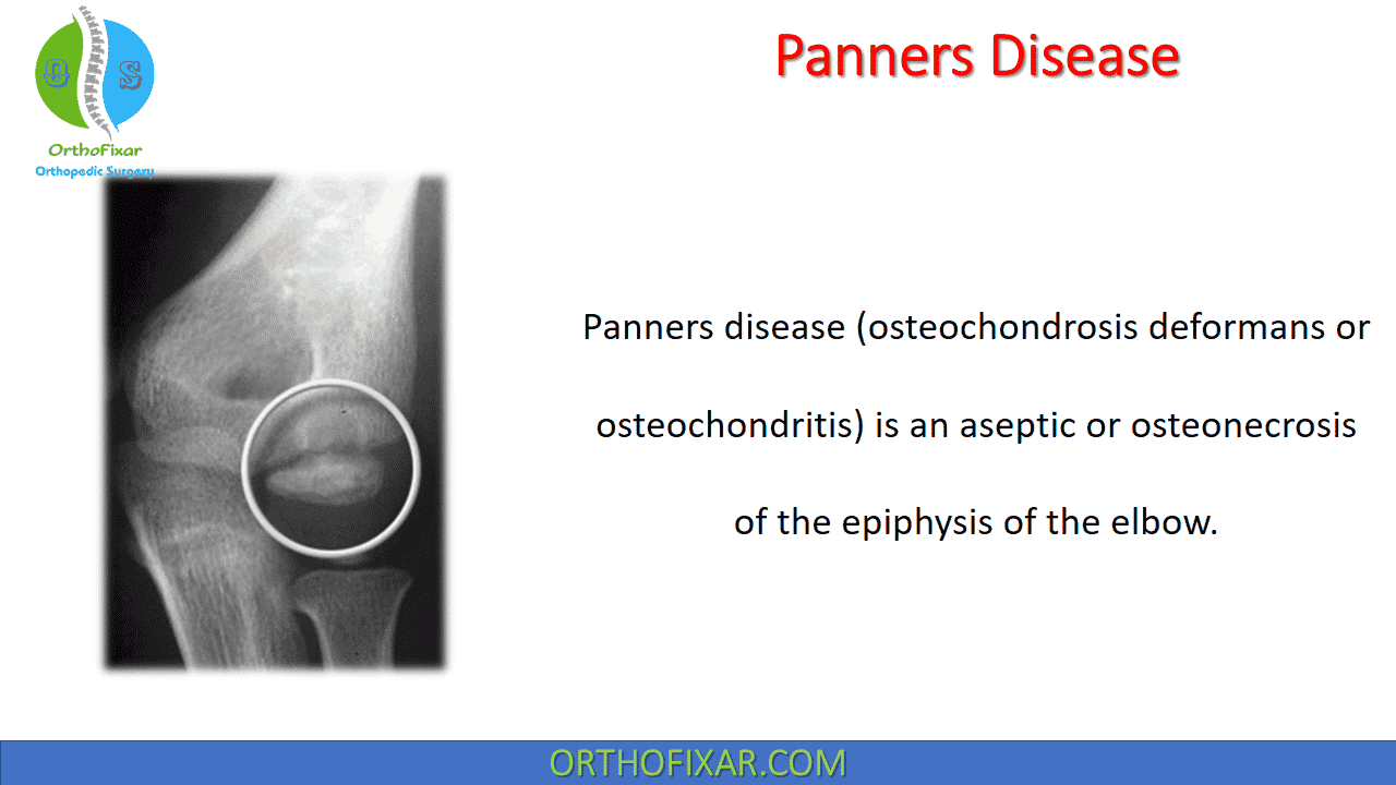
Panners disease (osteochondrosis deformans or osteochondritis) is an aseptic or osteonecrosis of the epiphysis of the elbow.
Panners Disease Causes
In 1927, Hans Jessen Panner published a description of osteochondrosis of the capitellum, likening it to Legg-Calvé-Perthes disease of the hip. Like other ostochondroses, it consists of noninflammatory, disordered endochondral ossification. Its specific cause and relation to OCD remain debatable.
It is known that abnormal radiocapitellar compressive forces during a period of vulnerability, typically while the physes are still open, predispose children to Panners disease. It may result from the combination of an avascular insult (likely related to the capitellum’s predominantly end-artery supply) and repetitive microtrauma.
Panner’s disease predominantly affects boys younger than age 10 years. Young boys may be predisposed to it for two reasons:
- First, compared with girls, they have a delayed appearance and maturation of their secondary growth centers.
- Second, boys traditionally are more prone to trauma during the more aggressive early childhood activities they select.
This may change as more girls choose higher-risk athletic activities at younger ages. Although Panners disease can be confused with OCD, and the age of onset may overlap, it is distinguished by three epidemiologic characteristics. Panners disease does not share the strict association with repetitive throwing that OCD does, it is usually self limited, and it resolves without any long-term sequelae.
See Also: Elbow Osteochondritis Dissecans

Clinical Evaluation
The main presenting symptoms are pain to the lateral aspect of the elbow, swelling, and a limitation of elbow movement in a noncapsular pattern.
If a displaced fragment is present, there is often a painless limitation of elbow extension, with a soft endfeel, but a hard end-feel when flexion is limited.
Imaging
Radiographs initially show fissuring, lucencies, fragmentation, and irregularity of the capitellum, particularly near or at the chondral surface.
Subsequent x-ray films, taken at 3 to 5 months, demonstrate larger radiolucent areas followed by reossification of the bony epiphysis, with a corresponding resolution of symptoms. In 1 to 2 years, the epiphysis regains its contour, usually without flattening. As in Legg Calvé Perthes, radiographs often lag behind clinical symptoms.
Magnetic resonance imaging (MRI) in Panners disease may be used effectively to document the extent of the lesion. Typically, edema is localized to the chondral surface and the bone adjacent to it, with less involvement of the deeper subchondral bone compared with the OCD
See Also: Legg Calvé Perthes Disease


Panner’s Disease Treatment
Panner’s Disease Treatment involves rest and cessation of the aggravating activity and administering therapeutic modalities such as ice and anti-inflammatory medication. The elbow occasionally may need to be immobilized for 3 to 4 weeks to control symptoms.
Symptoms usually resolve within 6 to 8 weeks, although they occasionally persist for months. Activities should be reinstituted progressively and as tolerated. The condition has an excellent long-term prognosis, although some patients may have a slight residual flexion contracture.
References
- Claessen FM, Louwerens JK, Doornberg JN, van Dijk CN, Eygendaal D, van den Bekerom MP. Panner’s disease: literature review and treatment recommendations. J Child Orthop. 2015 Feb;9(1):9-17. doi: 10.1007/s11832-015-0635-2. Epub 2015 Feb 7. PMID: 25663360; PMCID: PMC4340849.
- Douglas G, Rang M . The role of trauma in the pathogenesis of the osteochondroses. Clin Orthop Relat Res. 1981 ;( 158 ): 28 – 32 .
- Ruch DS, Poehling GG . Arthroscopic treatment of Panner’s disease . Clin Sports Med. 1991 ; 10 : 629 – 636 .
- Singer KM, Roy SP. Osteochondrosis of the humeral capitellum . Am J Sports Med. 1984 ; 12 : 351 – 360 .
- Panner H . An affection of the capitulum humeri resembling Calvé- Perthes disease of the hip . Acta Radiol. 1927 ; 8 : 617 – 618 .
- Duthie RB, Houghton GR . Constitutional aspects of the osteochondroses. Clin Orthop Relat Res. 1981 ;( 158 ): 19 – 27 .
- Yamaguchi K, Sweet FA, Bindra R , et al . The extraosseous and intraosseous arterial anatomy of the adult elbow. J Bone Joint Surg Am. 1997 ; 79 : 1653 – 1662 .
- Kobayashi K, Burton KJ, Rodner C , et al . Lateral compression injuries in the pediatric elbow: Panner’s disease and osteochondritis dissecans of the capitellum . J Am Acad Orthop Surg. 2004 ; 12 : 246 – 254 .
- Cyriax J: Textbook of Orthopaedic Medicine, Diagnosis of Soft Tissue Lesions, 8th ed. London: Bailliere Tindall, 1982
August 8, 2023
OrthoFixar
Orthofixar does not endorse any treatments, procedures, products, or physicians referenced herein. This information is provided as an educational service and is not intended to serve as medical advice.
- Lifetime product updates
- Install on one device
- Lifetime product support
- Lifetime product updates
- Install on one device
- Lifetime product support
- Lifetime product updates
- Install on one device
- Lifetime product support
- Lifetime product updates
- Install on one device
- Lifetime product support