Lateral Epicondylitis

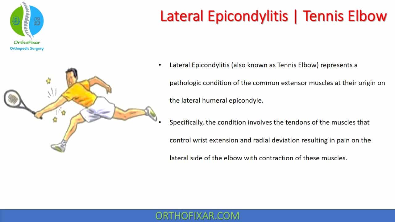
Lateral epicondylitis (Tennis Elbow) represents a pathologic condition of the common extensor muscles at their origin on the lateral humeral epicondyle.
Specifically, the condition involves the tendons of the muscles that control wrist extension and radial deviation resulting in pain on the lateral side of the elbow with contraction of these muscles.
While the terms epicondylitis and tendinitis are commonly used to describe tennis elbow, histopathologic studies have demonstrated that this condition is often not an inflammatory condition; rather, it is a degenerative condition, a tendinosis.
See Also: Elbow Anatomy
Historical Brief
The first description of tennis elbow is attributed to Runge, but the name derives from lawn tennis arm described by Morris in the Lancet in 1882. This was followed in 1883 by Dr H.P. Major describing his own affliction in 1883.
In Runge’s original description, he called it writer’s cramp and attributed it to a periostitis of the lateral humeral epicondyle. Since then, it has been referred to by a number of names including epicondylalgia, epicondyle pain, musician’s palsy, and tennis pain, although most authors have used the terms Lateral epicondylitis or tennis elbow.
Epidemiology
Tennis elbow affects between 1 and 3% of the population.
It occurs most commonly between 35 and 50 years of age with a mean age of 45, it is seldom seen in those under 20 years of age, .
Usually affects the dominant arm.
Cyriax noted that the origin of the ECRB was the primary site of this injury, and pathologic changes have been consistently documented at this location, although findings are also found in the ECRL, and the ECU. One-third of patients also have involvement of the origin of the EDC.

Lateral Epicondylitis Causes
Tennis elbow usually results from overuse although it can be traumatic in origin.
One possible etiology for overuse at the elbow is the fact that the hand does not have a supportive function but functions predominantly to grasp some object. Repetitive grasping, with the wrist positioned in extension, places the elbow particularly at risk. Participants of tennis, baseball, javelin, golf, squash, racquetball, swimming, and weight lifting are all predisposed to this risk.
Other conditions have been suggested as causes of tennis elbow, including
- periostitis:
- infection,
- bursitis,
- fibrillation of the radial head,
- radioulnar joint disease,
- Calcific tendinitis,
- neurogenic causes,
- osteochondritis dissecans,
- radial nerve entrapment,
- Capsular and ligamentous lesions,
See Also: Osteochondritis Dissecans
Dysfunction of the cervical spine has also been speculated to cause tennis elbow. Theoretically, a frank radiculopathy may weaken the extensor muscles to the point at which normal use is traumatic and induces a grade I tear in the muscle belly or tendon. Less definite compression of the nerve root may compromise axoplasmic transportation, producing a trophic malnutrition of the muscle, resulting in damage.
The shoulder has also been speculated to be a cause of tennis elbow, due to the effect of an abnormal tension in the clavipectoral fascia on the brachial plexus. It is speculated that if the fascia is distorted due to clavicular malposition, the traction exerted on the brachial plexus could lead to similar problems as those encountered with a cervical lesion.
Nirschl postulates that some patients who have tennis elbow may have a genetic predisposition that makes them more susceptible to tendinosis at multiple sites. He terms this condition mesenchymal syndrome on the basis of the stem-cell line of fibroblasts and the presence of a potentially systemic abnormality of cross-linkage in the collagen produced by the fibroblasts. Patients may have mesenchymal syndrome if they have two or more of the following conditions:
- Bilateral lateral tennis elbow
- Cubital tunnel syndrome
- CTS
- De Quervain tenosynovitis
- Trigger finger
- Rotator cuff tendinosis

Lateral Epicondylitis Stages
Nirschl previously categorized the stages of repetitive microtrauma:
Stage 1: injury is probably inflammatory, is not associated with pathologic alterations, and is likely to resolve.
Stage 2: injury is associated with pathologic alterations such as tendinosis or angiofibroblastic degeneration. It is this stage that is most commonly associated with sportsrelated tendon injuries such as tennis elbow, and with overuse injuries in general. Within the tendon, there is a fibroblastic and vascular response (tendinosis) rather than an immune blood-cell response (inflammation).
Stage 3: injury is associated with pathologic changes (tendinosis) and complete structural failure (rupture).
Stage 4: injury exhibits the features of a stage 2 or 3 injury and is associated with other changes such as fibrosis, soft matrix calcification, and hard osseous calcification. The changes that are associated with a stage 4 injury may also be related to the use of cortisone.
Clinical Presentation
Three types of tennis elbow are recognized based on the mode of onset:
- The acute onset (indirect) type of tennis elbow is associated with a recognizable mechanism with acute pain, associated bruising on occasion, and a feeling of something giving way within the elbow.
- Rupture of the ECRL with tenderness in the muscles is associated with direct trauma to the lateral side of the elbow, but with no tearing of the ligaments.
- The chronic type is associated with a gradual onset and is occasionally termed occupational neuralgia.
Tennis Elbow symptoms include:
- Pain: The pain of tennis elbow is often related to activities that involve wrist extension/grasp, as it is the wrist extensors that must contract during grasping activities to stabilize the wrist.
- Diffuse achiness.
- morning stiffness.
- Palpable tenderness is usually found over the ECRB and ECRL, especially at the lateral epicondyle, with the site of maximum tenderness most commonly being over the anterior aspect of the lateral epicondyle.
The next most common site is tenderness over the radial head, or where the lateral part of the common extensor tendon arises from the bone.
Occasionally the pain is experienced at night and the patient may report dropping objects frequently, especially if they are carried with the palm facing down.
Differentiation between the various tendons is obviously important. Five types of tendon lesions of the elbow are recognized:
- Type 1: a lesion of the muscle origin of the ECRL, which is usually located just proximal to the lateral epicondyle.
- Type 2: An insertion tendinopathy of the ECRB. This is the most common site and is usually associated with type 5.
- Type 3: As the ECRB also originates from the radial collateral ligament, involvement of the tendon can produce pain here, or at the radial head.
- Type 4: an ECRB muscle belly strain.
- Type 5: inflammation at the origin of the extensor digitorum.
The range-of-motion tests typically reveal the following:
- Active motions are usually painless, although there may be pain with wrist flexion when combined with elbow extension.
- Passive motion can produce pain, especially with passive wrist flexion with the forearm pronated and the elbow extended.
The resisted tests typically reproduce symptoms with resisted wrist extension and radial deviation with the elbow extended. Pain on resisted finger extension has also been reported.
Cozen’s test or Mills test are typically positive.
The cervical spine, shoulder, and wrist must also be examined. As a large number of tennis elbows appear to be secondary to a dysfunction of either the cervical spine or the shoulder, testing isometric wrist extension in varying positions of the cervical spine or shoulder will help differentiate the cause.
See Also: Elbow Examination


Lateral Epicondylitis Treatment
Tennis elbow is normally a self-limiting complaint; without intervention, the symptoms will usually resolve within 8–12 months.
Non-operative Treatment:
The initial treatment of tennis elbow should be conservative which include:
- avoidance of aggravating activities,
- anti-inflammatory drugs (NSAIDs),
- counterforce bracing,
- occupational therapy for local modalities (ice, heat, ultrasonography, iontophoresis),
- corticosteroid injection,
- Platelet-rich plasma (PRP) injection,
- Extracorporeal shock wave therapy.


Many different types of braces and other orthotic devices are available. The main type is a band or strap around the muscle belly of the wrist extensors. Theoretically, binding the muscle with a clasp, band, or brace should limit expansion and thereby decrease the contribution to force production by muscle fibers proximal to the band.
Counterforce bracing has been shown to beneficially impact force couple imbalances and altered movements, decrease angular acceleration at the elbow and decrease EMG activity.

Operative Treatment:
Tennis Elbow treatment with surgery is indicated if the symptoms do not resolve despite a properly performed conservative intervention lasting 6 months.
A simple handshake test can help to determine whether surgical treatment of tennis elbow is required. The patient is asked to perform a firm handshake with the elbow extended, and then supinate the forearm against resistance. The clinician notes whether the patient reports having pain at the origin of the extensors of the wrist. The elbow is then flexed to 90 degrees, and the same maneuver is performed. If pain is decreased in the flexed position, operative treatment is less likely to be needed. If the pain is equally severe with the elbow flexed and extended, then operative treatment is more likely to be needed.
The goals of operative treatment of Lateral Epicondylitis are to resect the pathologic material to stimulate neovascularization by producing focused local bleeding, and to create a healthy scar while doing the least possible structural damage to the surrounding tissues.
Surgical intervention may be open or arthroscopic( ECRB tendon débridement with or without lateral epicondylectomy).
Lateral Epicondylitis Surgery Procedures include:
- Boyd and McLeod procedure includes excision of the proximal portion of the annular ligament, release of the entire extensor origin, excision of an adventitious bursa (if found), and resection of hypertrophic synovium in the radiocapitellar articulation.
- Another procedure includes exposure of the diseased extensor carpi radialis brevis origin, resection of degenerative tissue, and direct repair to bone.
Postoperatively, a carefully guided resistance-based rehabilitation program is recommended.

Nirschl and Sobel have attempted to determine whether the presenting symptoms are helpful in both diagnosing and directing the intervention. This information was previously published in the form of a table:
| Type | Pain Description | Treatment |
|---|---|---|
| Type I | Pain is characterized by stiffness or mild soreness after activity and resolves within 24 hours. | usually self limiting when proper precautions are taken |
| Type II | Pain is marked by stiffness or mild soreness after exercise, lasts more than 48 hours, is relieved with warm-up exercises, is not present during activity, and resolves within 72 hours after the cessation of activity. The pain associated with types 1 and 2 may be due to peritendinous inflammation. | usually self limiting when proper precautions are taken |
| Type III | Pain is characterized by stiffness or mild soreness before activity and is partially relieved with warm-up exercises. The pain not prevent participation in activity and is only mild during activity. However, minor adjustments in the technique, intensity, and duration of activity are needed to control the pain. Type 3 pain may necessitate the use of nonsteroidal antiinflammatory medications. | usually respond to nonoperative medical therapy |
| Type IV | Pain is more intense than type 3 pain and produces changes in the performance of a specific sport- or work-related activity. Mild pain accompanies the activities of daily living. Type 4 pain may reflect tendon damage | usually respond to nonoperative medical therapy |
| Type V | Pain, which is characterized as moderate or severe before, during, and after exercise, greatly alters or prevents performance of the activity. Pain accompanies but does not prevent the performance of activities of daily living. Complete rest controls the pain. Type 5 pain reflects permanent tendon damage. | Operative treatment |
| Type VI | Pain, which is similar to type 5 pain, prevents the performance of activities of daily living and persists despite completve rest. | Operative treatment |
| Type VII | Pain is a consistent, aching pain that intensifies with activity and that regularly interrupts sleep. | Operative treatment |
References
- Field LD, Savoie FH. Common elbow injuries in sport. Sports Med. 1998 Sep;26(3):193-205. doi: 10.2165/00007256-199826030-00005. PMID: 9802175.
- Runge F: Zur genese und behandlung des schreibekrampfs. Berliner klinische Wochenschr :245–246, 1873.
- Morris H: The rider’s sprain. Lancet 29:133–134, 1882.
- Major HP: Lawn-tennis elbow. BMJ 15:557, 1883.
- Brandesky W: Uber den Epicondylusschmerz. Dtsch Zeitschr Chir 219:246–255, 1929.
- Nirschl RP: Prevention and treatment of elbow and shoulder injuries in the tennis player. Clin Sports Med 7:289–308, 1988.
- NirschlRP: Mesenchymal syndrome. Virginia Med Monthly 96:659–662, 1969.
- Nirschl RP: Patterns of failed tendon healing in tendon injury. In: Leadbetter WB, Buckwalter JA, Gordon SL, eds. Sports-Induced Inflammation: Clinical and Basic Science Concepts. Park Ridge, IL: American Academy of Orthopaedic Surgeons, 1990:609–618.
- Ernst E: Conservative therapy for tennis elbow. Br J Clin Pract 46:55–57, 1992.
- Nirschl RP, Pettrone FA: Tennis elbow. J Bone Joint Surg [Am] 61- A:832–839, 1979.
- Nirschl RP, Sobel J: Arm Care. A Complete Guide to Prevention and Treatment of Tennis Elbow. Arlington, VA: Medical Sports, 1996.
- Kraushaar BS, Nirschl RP: Pearls: handshake lends epicondylitis cues. Phys Sportsmed 24:15, 1996.
- Lee DG: “Tennis elbow”: A manual therapist’s perspective. J Orthop Sports Phys Ther 8:134–142, 1986.
- Nirschl RP: Elbow tendinosis: tennis elbow. Clin Sports Med 11:851–870, 1992.
- Nirschl RP: Muscle and tendon trauma: tennis elbow. In: Morrey BF, ed. The Elbow and Its Disorders, 2nd ed. Philadelphia, PA: WB Saunders, 1993:681–703.
- Kraushaar BS, Nirschl RP. Tendinosis of the elbow (tennis elbow). Clinical features and findings of histological, immunohistochemical, and electron microscopy studies. J Bone Joint Surg Am. 1999 Feb;81(2):259-78. PMID: 10073590.
- Cyriax JH: The pathology and treatment of tennis elbow. J Bone Joint Surg 18:921–940, 1936.
- Nirschl RP: Tennis elbow tendinosis: pathoanatomy, nonsurgical and surgical management. In: Gordon SL, Blair SJ, Fine LJ, eds. Repetitive Motion Disorders of the Upper Extremity. Rosemont, IL: American Academy of Orthopaedic Surgeons, 1995:467–479.
- Mills PG: The treatment of , tennis elbow,. BMJ 1:12–13, 1928.
- Kaplan EB: Treatment of tennis elbow (epicondylitis) by denervation. J Bone Joint Surg Am 41A:147–151, 1959.
- Bosworth DM: Surgical treatment of tennis elbow: a followup study. J Bone Joint Surg 47A:1533–1536, 1965.
- Gunn C, Milbrandt W: Tennis elbow and the cervical spine. Can Med Assoc J 114:803–809, 1976.
- Groppel J, Nirschl RP: A biomechanical and electromyographical analysis of the effects of counter force braces on the tennis player. Am J Sports Med 14:195–200, 1986.
- Krischek O, Hopf C, Nafe B, et al: Shock-wave therapy for tennis and golfer’s elbow–1 year follow-up. Arch Orthop Trauma Surg 119:62–66, 1999.
- Lahz JRS: Concerning the pathology and treatment of tennis elbow. Med J Aust 2:737–742, 1947.
- Dutton’s Orthopaedic Examination, Evaluation, And Intervention 3rd Edition.
- Campbel’s Operative Orthopaedics 12th edition Book.
June 11, 2023
OrthoFixar
Orthofixar does not endorse any treatments, procedures, products, or physicians referenced herein. This information is provided as an educational service and is not intended to serve as medical advice.
- Lifetime product updates
- Install on one device
- Lifetime product support
- Lifetime product updates
- Install on one device
- Lifetime product support
- Lifetime product updates
- Install on one device
- Lifetime product support
- Lifetime product updates
- Install on one device
- Lifetime product support