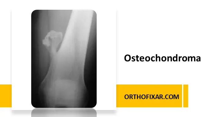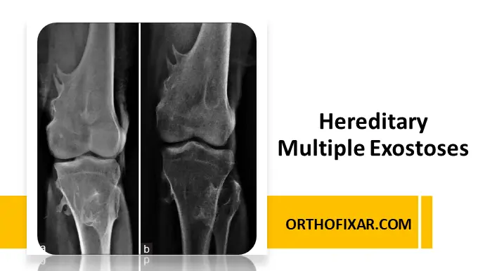
Hereditary multiple exostoses (HME), also known as hereditary multiple osteochondromas (HMO), is a rare autosomal dominant skeletal disorder characterized by the development of multiple cartilage-capped bony growths called osteochondromas. These benign tumors usually arise near the growth plates of long bones during childhood and adolescence.
Although osteochondromas are benign, HME can lead to skeletal deformities, chronic pain, restricted joint movement, limb-length discrepancies, and, in rare cases, malignant transformation into chondrosarcoma.
This article provides an evidence-based medical overview of hereditary multiple exostoses, including causes, symptoms, diagnosis, treatment, prognosis, and recent research developments.
What is Hereditary Multiple Exostoses?
Hereditary multiple exostoses is a genetic disorder affecting bone growth. Patients develop multiple osteochondromas, which are abnormal bony projections covered with cartilage.
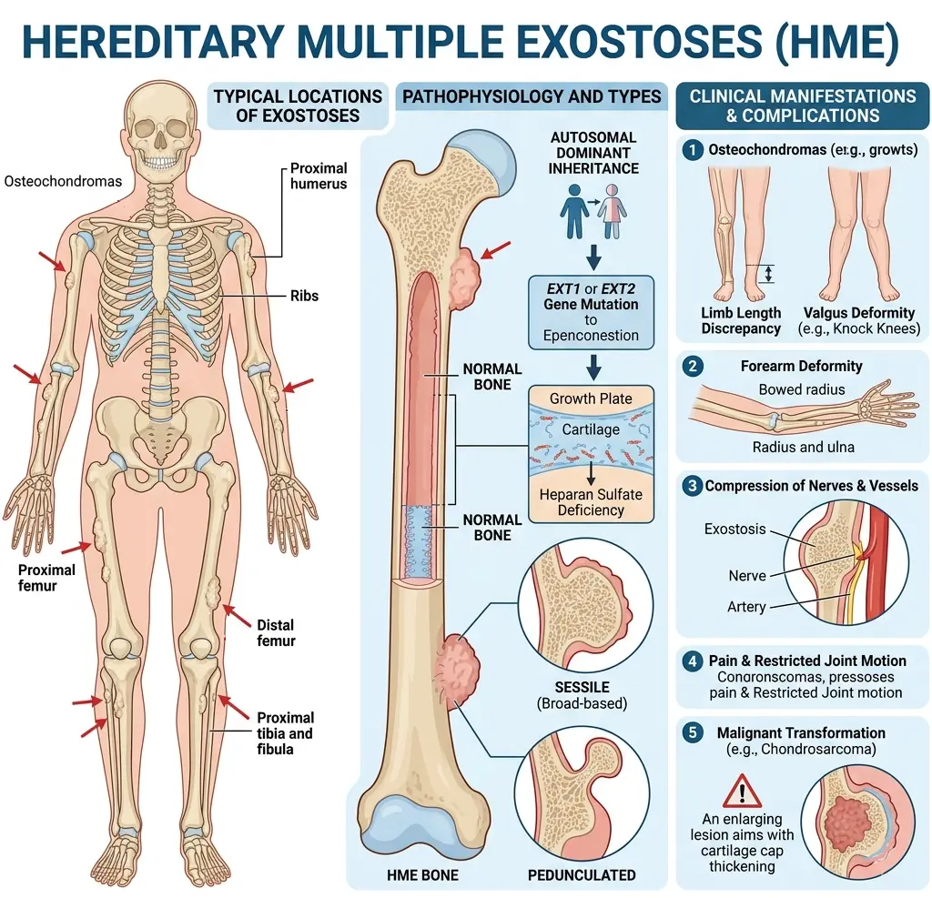
These growths commonly occur in:
- Femur
- Tibia
- Humerus
- Forearm bones
- Pelvis
- Ribs
- Scapula
The condition typically presents in early childhood, with most patients diagnosed before age 12.
Epidemiology of HME
HME is considered a rare disease with an estimated prevalence of approximately 1 in 50,000 individuals worldwide. Males are slightly more frequently affected than females.
The disorder demonstrates high penetrance, meaning most individuals carrying the pathogenic mutation eventually develop clinical manifestations.
Causes and Genetics of Hereditary Multiple Exostoses
EXT1 and EXT2 Gene Mutations
Most cases of hereditary multiple exostoses are caused by mutations in the EXT1 and EXT2 genes.
These genes are responsible for producing enzymes involved in heparan sulfate synthesis, an essential component in cartilage growth and skeletal development.
The disease follows an autosomal dominant inheritance pattern, meaning:
- One mutated copy of the gene is sufficient to cause disease
- Each child of an affected parent has a 50% chance of inheriting the condition
Research shows:
- EXT1 mutations are often associated with more severe disease
- EXT2 mutations may produce milder phenotypes
Pathophysiology
The defective EXT genes impair normal heparan sulfate biosynthesis. This disrupts signaling pathways involved in chondrocyte proliferation and growth plate organization.
As a result, cartilage cells grow abnormally outward from the metaphysis of bones, forming osteochondromas.
See Also: Bone Formation & Development
Symptoms of Hereditary Multiple Exostoses
Clinical manifestations vary considerably between individuals.
Common Symptoms
Multiple Bone Growths
Patients develop hard, palpable masses near joints and long bones.
Skeletal Deformities
Common deformities include:
- Bowing of the forearm
- Short stature
- Knee valgus deformity
- Limb-length discrepancies
- Coxa valga
Pain
Pain may result from:
- Mechanical irritation
- Compression of surrounding tissues
- Bursa formation
- Joint dysfunction
Restricted Movement
Large osteochondromas may limit joint mobility and impair physical activity.
Neurovascular Compression
Some lesions compress nerves or blood vessels, causing:
- Numbness
- Tingling
- Weakness
- Vascular insufficiency
Complications of HME
Malignant Transformation
The most serious complication is transformation into secondary peripheral chondrosarcoma.
Warning signs include:
- Sudden enlargement of a lesion after skeletal maturity
- Increasing pain
- Thickened cartilage cap
- Neurological symptoms
The estimated lifetime risk of malignant transformation ranges from approximately 1% to 5%.
Orthopedic Complications
Patients may develop:
- Premature osteoarthritis
- Fractures
- Spinal cord compression
- Chronic disability
Diagnosis of Hereditary Multiple Exostoses
Clinical Evaluation
Diagnosis begins with:
- Family history
- Physical examination
- Assessment of skeletal deformities
Imaging Studies
X-rays
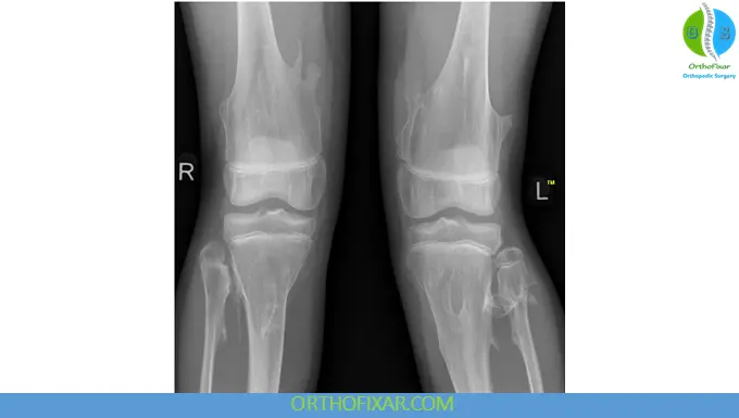
Radiographs remain the primary imaging modality and reveal characteristic osteochondromas near growth plates.
MRI
MRI is useful for:
- Evaluating cartilage cap thickness
- Detecting malignant transformation
- Assessing neurovascular involvement
CT Scan
CT imaging helps evaluate complex anatomical regions such as the pelvis or spine.
Genetic Testing
Molecular testing can identify pathogenic variants in:
- EXT1
- EXT2
Genetic testing confirms diagnosis and supports family counseling.
Differential Diagnosis
Conditions that may resemble HME include:
- Solitary osteochondroma
- Metachondromatosis
- Ollier disease
- Langer-Giedion syndrome
- Trevor disease
Treatment of Hereditary Multiple Exostoses
There is currently no cure for HME. Treatment focuses on symptom management and complication prevention.
Conservative Management
Mild cases may only require:
- Observation
- Periodic imaging
- Physical therapy
- Pain management
Surgical Treatment
Surgery may be indicated for:
- Painful lesions
- Functional impairment
- Neurovascular compression
- Severe deformity
- Suspicion of malignancy
Procedures include:
- Osteochondroma excision
- Corrective osteotomy
- Limb-length correction
Long-Term Monitoring
Regular follow-up is essential to monitor:
- Skeletal development
- Tumor growth
- Malignant transformation
Prognosis
Most patients with hereditary multiple exostoses maintain a normal lifespan. However, quality of life may be affected by chronic pain, repeated surgeries, and skeletal deformities.
Early diagnosis and multidisciplinary management significantly improve functional outcomes.
Genetic Counseling
Because HME is inherited in an autosomal dominant pattern, genetic counseling is strongly recommended for affected families.
Prenatal and preimplantation genetic testing may be considered in some cases.
Recent Research and Future Therapies
Current research focuses on:
- Molecular pathways involving heparan sulfate synthesis
- Targeted therapies for abnormal cartilage growth
- Improved genetic diagnostics
- Novel treatments to prevent osteochondroma formation
Experimental therapies are under investigation but are not yet clinically available.
Conclusion
Hereditary multiple exostoses is a rare genetic skeletal disorder characterized by multiple osteochondromas and progressive orthopedic complications. Mutations in EXT1 and EXT2 disrupt normal bone growth and lead to abnormal cartilage proliferation.
Early recognition, regular monitoring, imaging studies, and individualized orthopedic care are essential for optimal outcomes. Although no definitive cure exists, advances in molecular genetics continue to improve understanding and management of this complex disorder.
Frequently Asked Questions (FAQs)
Is hereditary multiple exostoses cancerous?
HME itself is benign, but a small percentage of patients may develop chondrosarcoma later in life.
At what age is HME diagnosed?
Most patients are diagnosed during childhood, typically before age 12.
Can hereditary multiple exostoses skip generations?
Because HME is autosomal dominant with high penetrance, it usually does not skip generations, although symptom severity can vary.
Is there a cure for HME?
Currently, no cure exists. Treatment focuses on symptom management and surgical correction when needed.
Which genes are involved in HME?
The condition is most commonly associated with mutations in the EXT1 and EXT2 genes.
References & More
- Sefcik R, Earl D, Thorpe S. Hereditary Multiple Osteochondromas. 2000 Aug 3 [Updated 2026 Jan 29]. In: Adam MP, Bick S, Mirzaa GM, et al., editors. GeneReviews® [Internet]. Seattle (WA): University of Washington, Seattle; 1993-2026. Available from: https://www.ncbi.nlm.nih.gov/books/NBK1235/
- Rueda-de-Eusebio A, Gomez-Pena S, Moreno-Casado MJ, Marquina G, Arrazola J, Crespo-Rodríguez AM. Hereditary multiple exostoses: an educational review. Insights Imaging. 2025 Feb 21;16(1):46. doi: 10.1186/s13244-025-01899-6. PMID: 39982564; PMCID: PMC11845651. Pubmed
- Beltrami G, Ristori G, Scoccianti G, Tamburini A, Capanna R. Hereditary Multiple Exostoses: a review of clinical appearance and metabolic pattern. Clin Cases Miner Bone Metab. 2016 May-Aug;13(2):110-118. doi: 10.11138/ccmbm/2016.13.2.110. Epub 2016 Oct 5. PMID: 27920806; PMCID: PMC5119707. Pubmed