Mucopolysaccharidoses (MPS) are a group of rare inherited lysosomal storage disorders caused by deficiencies in enzymes responsible for breaking down glycosaminoglycans (GAGs). The progressive accumulation of these complex carbohydrates in cells and tissues produces multisystem disease, particularly affecting the skeleton, joints, nervous system, cardiovascular system, respiratory tract, liver, and spleen.
The major mucopolysaccharidosis types include Hurler syndrome (MPS I), Hunter syndrome (MPS II), Sanfilippo syndrome (MPS III), Morquio syndrome (MPS IV), Maroteaux–Lamy syndrome (MPS VI), and Sly syndrome (MPS VII).
Clinical manifestations range from severe childhood-onset neurodegeneration to predominantly skeletal abnormalities with preserved intellectual development. Early recognition is essential because several MPS types have disease-specific treatments that may slow progression and improve clinical outcomes.
This medical education review explains the causes, classification, inheritance patterns, clinical manifestations, skeletal abnormalities, diagnosis, treatment, and prognosis of mucopolysaccharidoses. [1]
What Are Mucopolysaccharidoses (MPS)?
Mucopolysaccharidoses are inherited metabolic diseases belonging to the broader category of lysosomal storage disorders.
Lysosomes are intracellular organelles containing enzymes that degrade complex molecules. In patients with MPS, a specific lysosomal enzyme is absent or has markedly reduced activity, resulting in incomplete degradation of glycosaminoglycans.
Glycosaminoglycans, previously called mucopolysaccharides, are long, unbranched polysaccharides that form important components of the extracellular matrix.
They are particularly abundant in:
- Bone and cartilage
- Ligaments and tendons
- Skin and connective tissues
- Cornea
- Blood vessel walls and cardiac valves
- Joint structures
- Various internal organs
When GAGs cannot be adequately degraded, they accumulate within lysosomes, producing progressive cellular dysfunction, inflammation, tissue damage, and organ impairment.
Major Glycosaminoglycans Involved in MPS
The principal accumulated GAGs include:
- Heparan sulfate: Particularly associated with neurological involvement.
- Dermatan sulfate: Accumulates in connective tissues, skin, cardiac valves, and other organs.
- Keratan sulfate: Prominent in cartilage, bone, and corneal tissues.
- Chondroitin sulfate: Important in cartilage and skeletal tissues.
- Hyaluronan (hyaluronic acid): Accumulates in the exceptionally rare MPS IX.
The combination of accumulated GAGs varies according to the deficient enzyme and helps explain differences between MPS subtypes. [1]
Epidemiology and Prevalence of Mucopolysaccharidoses
Mucopolysaccharidoses are rare diseases, but their combined frequency is clinically significant.
Some published reviews estimate an overall birth incidence of approximately 1 in 25,000 live births, although reported rates vary considerably by geographic region, population, diagnostic practices, and study methodology.
A United States retrospective study covering diagnoses between 1995 and 2015 reported:
- Birth incidence of approximately 0.98 cases per 100,000 live births.
- Estimated prevalence of 2.67 cases per million population.
- MPS I, II, and III among the most frequently identified types.
These findings illustrate why a single worldwide prevalence estimate should not be applied to every population.
Importantly, Hurler syndrome and Sanfilippo syndrome are relatively common among MPS disorders, but the most frequently reported subtype differs between countries. [6]
Causes and Genetic Inheritance of Mucopolysaccharidoses
Mucopolysaccharidoses result from pathogenic genetic variants affecting lysosomal enzymes involved in GAG degradation.
Autosomal Recessive Inheritance
Most MPS types follow an autosomal recessive inheritance pattern.
These include MPS I, III, IV, VI, VII, and IX.
For two carrier parents, each pregnancy has:
- A 25% probability of producing an affected child.
- A 50% probability of producing an unaffected carrier.
- A 25% probability of producing a child who is neither affected nor a carrier of the familial variants.
Males and females can be affected.
X-Linked Inheritance
Hunter syndrome (MPS II) is inherited in an X-linked pattern because its causative gene, IDS, is located on the X chromosome.
The disorder predominantly affects males. However, rare affected females have been documented, so it is inaccurate to state that Hunter syndrome occurs exclusively in males.
Molecular genetic testing and genetic counseling are important for affected families. [2,3]
Classification and Types of Mucopolysaccharidoses
MPS classification is based on the specific deficient lysosomal enzyme, the corresponding gene, and the accumulated glycosaminoglycans.
Classification of MPS by Enzyme Deficiency
| MPS type | Syndrome | Deficient enzyme | Accumulated GAGs |
|---|---|---|---|
| MPS I | Hurler / attenuated MPS I | α-L-iduronidase | Heparan sulfate, dermatan sulfate |
| MPS II | Hunter | Iduronate-2-sulfatase | Heparan sulfate, dermatan sulfate |
| MPS IIIA | Sanfilippo A | Heparan-N-sulfatase | Heparan sulfate |
| MPS IIIB | Sanfilippo B | α-N-acetylglucosaminidase | Heparan sulfate |
| MPS IIIC | Sanfilippo C | Heparan-α-glucosaminide N-acetyltransferase | Heparan sulfate |
| MPS IIID | Sanfilippo D | N-acetylglucosamine-6-sulfatase | Heparan sulfate |
| MPS IVA | Morquio A | N-acetylgalactosamine-6-sulfatase | Keratan sulfate, chondroitin-6-sulfate |
| MPS IVB | Morquio B | β-galactosidase | Keratan sulfate |
| MPS VI | Maroteaux–Lamy | Arylsulfatase B | Dermatan sulfate |
| MPS VII | Sly | β-glucuronidase | Multiple GAGs |
| MPS IX | Hyaluronidase deficiency | Hyaluronidase 1 | Hyaluronan |
Important classification note: Scheie syndrome was historically designated MPS V, but it is now recognized as part of the attenuated MPS I spectrum rather than a separate MPS type.
MPS numbering is historical and does not represent a simple sequence of disease severity. [1,11]
Clinical Features of Mucopolysaccharidoses
Although clinical presentation differs among subtypes, many patients develop recognizable multisystem manifestations.
General Clinical Manifestations
Common features include:
- Progressive short stature or growth failure
- Disproportionate skeletal growth
- Coarse facial features
- Macrocephaly
- Macroglossia
- Thickened skin and soft tissues
- Hepatomegaly and splenomegaly
- Recurrent respiratory infections
- Umbilical or inguinal hernias
- Joint stiffness or contractures
- Hearing impairment
- Corneal clouding in selected MPS types
- Cardiac valvular abnormalities
- Spinal deformity or instability
Developmental delay, intellectual disability, or neurological regression may occur, particularly in severe MPS I, neuronopathic MPS II, and MPS III.
Not all MPS patients have intellectual disability. Cognitive function is often preserved in Morquio syndrome, Maroteaux–Lamy syndrome, and attenuated forms of MPS I.
Growth Abnormalities
Affected children may have initially normal growth followed by slowing of linear growth during early childhood.
Disproportionate short stature may result from:
- Vertebral abnormalities
- Abnormal cartilage development
- Hip dysplasia
- Joint contractures
- Progressive skeletal deformity
Some patients have a particularly short trunk because of severe vertebral involvement.
Skeletal and Orthopedic Manifestations of MPS
Musculoskeletal abnormalities are among the most important diagnostic clues.
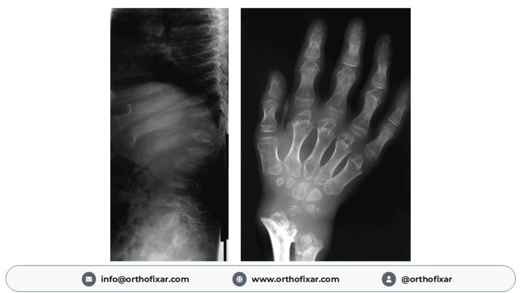
The characteristic radiographic pattern is called dysostosis multiplex, a collection of skeletal abnormalities resulting from abnormal bone and cartilage development.
Dysostosis Multiplex
Typical radiographic findings include:
- Abnormally shaped or flattened vertebral bodies
- Anterior vertebral beaking
- Thoracolumbar kyphosis
- Broad or paddle-shaped ribs
- Thickened clavicles
- Abnormal pelvis and acetabular development
- Hip dysplasia
- Short and broad metacarpals
- Proximal pointing of metacarpal bones
- J-shaped sella turcica in some patients
The severity and distribution of dysostosis multiplex vary by MPS subtype.
Spinal Abnormalities
Spinal involvement may include:
Thoracolumbar Kyphosis and Gibbus Deformity
Anterior vertebral wedging can produce a localized angular kyphosis known as a gibbus deformity.
This is particularly characteristic of severe MPS I but also occurs in other subtypes.
Atlantoaxial Instability
Odontoid hypoplasia, abnormal ligamentous structures, and GAG deposition around the cervical spine may produce atlantoaxial instability or spinal canal narrowing.
Cervical spinal cord compression is a serious complication, particularly in Morquio syndrome.
Progressive myelopathy may cause:
- Gait deterioration
- Weakness
- Sensory disturbances
- Abnormal reflexes
- Loss of bladder or bowel control
- Paralysis in advanced cases
Kyphoscoliosis and Spinal Stenosis
Progressive spinal deformity may impair sitting balance, walking, respiratory mechanics, or neurological function.
Hip, Knee, and Foot Abnormalities
Orthopedic manifestations include:
- Coxa valga
- Hip dysplasia
- Femoral head abnormalities
- Progressive hip arthritis
- Genu valgum
- Joint contractures
- Abnormal foot alignment
- Impaired standing and walking
A notable exception to the usual joint-stiffness pattern is Morquio syndrome, in which ligamentous laxity and joint hypermobility may coexist with skeletal deformity and pain.
Early orthopedic evaluation is important to prevent irreversible functional deterioration. [5,9]
Hurler Syndrome (MPS Type I)
Hurler syndrome represents the severe end of the MPS I clinical spectrum.
Cause and Enzyme Deficiency
MPS I results from deficient α-L-iduronidase, encoded by the IDUA gene.
This leads to accumulation of:
- Heparan sulfate
- Dermatan sulfate
MPS I has historically been divided into:
- Hurler syndrome — severe MPS I.
- Hurler–Scheie syndrome — intermediate phenotype.
- Scheie syndrome — attenuated phenotype.
Modern clinical classification generally distinguishes severe MPS I from attenuated MPS I because clinical manifestations overlap.
Clinical Manifestations of Hurler Syndrome
Children with severe MPS I may appear relatively normal at birth, with manifestations becoming evident during infancy or early childhood.
Typical features include:
- Progressive coarse facial features
- Macrocephaly and macroglossia
- Hepatosplenomegaly
- Corneal clouding
- Recurrent respiratory infections
- Hearing impairment
- Umbilical or inguinal hernias
- Progressive skeletal deformities
- Joint stiffness
- Developmental delay and subsequent cognitive decline
Growth and developmental progression commonly slow during the first years of life.
Orthopedic Features of MPS I
Characteristic abnormalities include:
- Thoracolumbar gibbus deformity
- Anterior vertebral beaking
- Odontoid hypoplasia
- Hip dysplasia
- Coxa valga
- Genu valgum
- Progressive joint contractures
- Carpal tunnel syndrome
Prognosis and Treatment
Untreated severe MPS I can lead to major cardiopulmonary and neurological complications and premature death.
Early hematopoietic stem cell transplantation (HSCT), ideally before substantial neurological impairment, is considered standard treatment for eligible young children with severe MPS I.
Enzyme replacement therapy with laronidase improves several non-neurological manifestations but does not adequately cross the blood–brain barrier.
Early diagnosis substantially affects long-term treatment outcomes. [2]
Hunter Syndrome (MPS Type II)
Hunter syndrome is an X-linked lysosomal storage disorder caused by deficiency of iduronate-2-sulfatase, encoded by the IDS gene.
It results in the accumulation of heparan sulfate and dermatan sulfate.
Clinical Features
Hunter syndrome shares many manifestations with Hurler syndrome, including:
- Coarse facial features
- Macrocephaly
- Macroglossia
- Hepatosplenomegaly
- Short stature
- Joint stiffness and contractures
- Hearing impairment
- Recurrent respiratory infections
- Cardiac disease
- Skeletal abnormalities
Unlike MPS I, significant corneal clouding is generally absent in Hunter syndrome, making this a useful clinical distinction.
Neurological Involvement
Hunter syndrome includes two broad clinical patterns:
Neuronopathic MPS II: Progressive neurocognitive impairment accompanied by systemic complications.
Non-neuronopathic MPS II: Predominantly systemic disease with relatively preserved intellectual function.
Disease severity and life expectancy vary substantially.
Musculoskeletal Manifestations
Joint contractures may begin during early childhood.
Progressive involvement can produce:
- Restricted shoulder movement
- Limited elbow extension
- Finger stiffness
- Hip abnormalities
- Carpal tunnel syndrome
- Progressive gait impairment
Treatment
Conventional enzyme replacement therapy with idursulfase can improve systemic manifestations but does not adequately treat neurological disease.
A significant development occurred in March 2026 when the United States granted accelerated approval to tividenofusp alfa-eknm (Avlayah), a brain-penetrant enzyme replacement therapy, for neurological manifestations of MPS II in eligible pediatric patients weighing at least 5 kg, before advanced neurological impairment.
This accelerated approval was based on reduction of cerebrospinal fluid heparan sulfate, a biomarker of disease activity. Continued approval may depend on confirmation of clinical benefit.
Therefore, the evidence should not be interpreted as proof that established neurological impairment can be reversed. [3,10]
Sanfilippo Syndrome (MPS Type III)
Sanfilippo syndrome is predominantly a neurodegenerative form of mucopolysaccharidosis caused by deficient degradation of heparan sulfate.
Unlike many other MPS disorders, severe neurological involvement is often more prominent than skeletal abnormalities.
Types of Sanfilippo Syndrome
Four established subtypes are recognized:
- MPS IIIA: Heparan-N-sulfatase deficiency, associated with SGSH.
- MPS IIIB: α-N-acetylglucosaminidase deficiency, associated with NAGLU.
- MPS IIIC: Heparan-α-glucosaminide N-acetyltransferase deficiency, associated with HGSNAT.
- MPS IIID: N-acetylglucosamine-6-sulfatase deficiency, associated with GNS.
Clinical Features
Early development may initially appear typical.
Symptoms commonly become recognizable during early childhood and may include:
- Speech delay
- Developmental delay
- Hyperactivity
- Behavioral disturbances
- Sleep abnormalities
- Progressive intellectual impairment
- Loss of previously acquired skills
- Seizures
- Progressive difficulties with movement and swallowing
As neurological deterioration progresses, affected individuals may lose independent walking ability and require mobility assistance.
Skeletal Manifestations
Orthopedic abnormalities are usually less prominent than those seen in MPS I, IV, or VI.
Possible findings include:
- Mild dysostosis multiplex
- Joint stiffness
- Scoliosis
- Hip abnormalities
- Progressive mobility limitations
Treatment and Prognosis
There is currently no established approved disease-modifying treatment for MPS III.
Management focuses on neurological symptoms, behavioral difficulties, sleep disturbance, seizure control, nutritional support, rehabilitation, and family-centered care.
Gene therapy and other approaches are being investigated.
Disease progression is highly variable; severe childhood-onset forms may cause death during childhood or adolescence, while attenuated forms can permit survival into adulthood. [4]
Morquio Syndrome (MPS Type IV)
Morquio syndrome, also called Morquio–Brailsford syndrome, predominantly affects skeletal development.
It is divided into two distinct biochemical subtypes.
MPS IVA (Morquio A)
MPS IVA results from deficiency of N-acetylgalactosamine-6-sulfatase, encoded by GALNS.
It causes accumulation of:
- Keratan sulfate
- Chondroitin-6-sulfate
MPS IVB (Morquio B)
MPS IVB results from deficiency of β-galactosidase, encoded by GLB1, and is associated primarily with keratan sulfate accumulation.
Clinical Manifestations
Morquio syndrome is characterized by disproportionate skeletal abnormalities, often with preserved intellectual development.
Typical features include:
- Severe short stature
- Short trunk and short neck
- Pectus carinatum
- Barrel-shaped chest
- Thoracolumbar kyphosis
- Scoliosis
- Genu valgum
- Hip dysplasia
- Joint hypermobility
- Progressive gait abnormalities
Facial coarsening is often less pronounced than in Hurler syndrome.
Cervical Spine Involvement
One of the most dangerous complications of MPS IV is cervical spinal cord compression.
Odontoid hypoplasia and atlantoaxial instability may place patients at risk of neurological injury.
Regular specialist evaluation and appropriate cervical spine imaging are essential.
Treatment
Elosulfase alfa is an enzyme replacement therapy available for MPS IVA.
It can improve endurance and certain systemic manifestations, although its ability to reverse established skeletal deformities is limited.
There is no equivalent established enzyme replacement therapy for MPS IVB.
Orthopedic procedures may be required to address spinal instability, hip deformities, knee malalignment, and other function-limiting complications.
Patients with attenuated disease may survive well into adulthood, although severe disease can substantially reduce life expectancy. [5]
Scheie Syndrome and Attenuated MPS I
Scheie syndrome was historically classified as MPS V.
It is now recognized as a relatively mild presentation within the attenuated MPS I spectrum.
Affected individuals may have:
- Joint stiffness
- Reduced hand mobility
- Carpal tunnel syndrome
- Corneal clouding
- Cardiac valve disease
- Skeletal abnormalities
- Relatively preserved intellectual development
Compared with untreated severe Hurler syndrome, manifestations often progress more slowly.
Nevertheless, attenuated MPS I can produce serious cardiac, respiratory, ophthalmological, and musculoskeletal complications.
Treatment may include laronidase and multidisciplinary supportive care. [2]
Maroteaux–Lamy Syndrome (MPS Type VI)
Maroteaux–Lamy syndrome results from deficient arylsulfatase B, also known as N-acetylgalactosamine-4-sulfatase.
The responsible gene is ARSB.
This defect predominantly causes dermatan sulfate accumulation.
Clinical Features
MPS VI resembles MPS I in several systemic and skeletal manifestations.
Common findings include:
- Short stature
- Coarse facial features
- Corneal clouding
- Hepatosplenomegaly
- Joint stiffness
- Hip dysplasia
- Thoracolumbar kyphosis
- Cardiac valve disease
- Respiratory compromise
- Hearing impairment
Intellectual Development
Primary intellectual development is typically preserved in MPS VI.
However, normal intellectual development does not imply mild systemic disease. Severe MPS VI can produce major skeletal, cardiopulmonary, and visual complications.
Treatment
Enzyme replacement therapy with galsulfase is recommended after confirmed diagnosis in appropriate patients.
Treatment can improve endurance and other systemic manifestations, but established skeletal abnormalities may continue to require orthopedic intervention.
Clinical severity and life expectancy vary, with many treated patients surviving into adulthood. [7]
Sly Syndrome (MPS Type VII)
Sly syndrome is caused by deficiency of β-glucuronidase, encoded by GUSB.
This leads to accumulation of several GAGs, including dermatan sulfate, heparan sulfate, and chondroitin sulfates.
Clinical Features
Clinical presentation is highly variable.
Manifestations may include:
- Nonimmune hydrops fetalis
- Hepatosplenomegaly
- Short stature
- Coarse facial features
- Skeletal abnormalities
- Corneal clouding
- Developmental delay
- Cardiac and respiratory complications
Nonimmune hydrops fetalis is an especially important diagnostic clue.
Treatment
Vestronidase alfa-vjbk is an enzyme replacement therapy available for MPS VII.
It addresses certain non-neurological manifestations but does not reliably correct established CNS or skeletal disease.
Patients require individualized, multidisciplinary follow-up. [8]
Mucopolysaccharidosis Type IX
MPS IX is an exceptionally rare lysosomal storage disorder caused by deficiency of hyaluronidase 1, encoded by HYAL1.
The principal accumulated substance is hyaluronan.
Reported manifestations have included:
- Joint swelling
- Synovial abnormalities
- Arthropathy
- Joint pain and reduced mobility
Unlike many other MPS types, prominent joint disease may occur without the usual severe multisystem phenotype.
Because so few patients have been reported, the full clinical spectrum remains uncertain. [12]
Diagnosis of Mucopolysaccharidoses
Early and accurate diagnosis is essential for identifying the MPS subtype, predicting complications, guiding therapy, and providing genetic counseling.
1. Clinical Examination
MPS should be considered in a child with a combination of unexplained:
- Short stature
- Progressive skeletal deformity
- Coarse facial features
- Joint contractures
- Organomegaly
- Corneal clouding
- Recurrent ear or respiratory disease
- Developmental delay or regression
Clinical examination should include assessment of growth, neurological function, joint mobility, vision, hearing, cardiac status, and respiratory function.
2. Urinary Glycosaminoglycan Testing
Urinary GAG analysis is a useful screening investigation.
Quantitative testing measures total or individual GAG concentrations, while qualitative methods identify characteristic GAG patterns.
However, normal urinary GAG testing does not reliably exclude all MPS disorders, particularly some attenuated phenotypes and MPS IV.
Urine testing alone is insufficient to establish the precise enzyme deficiency.
3. Enzyme Activity Assays
Definitive biochemical evaluation involves measurement of the relevant lysosomal enzyme activity.
Samples may include:
- Leukocytes
- Cultured fibroblasts
- Dried blood spots for selected assays
A deficient enzyme result must be interpreted alongside clinical findings and, where necessary, GAG and genetic testing.
For MPS I, pseudodeficiency can complicate interpretation of enzyme activity alone.
4. Molecular Genetic Testing
Molecular genetic analysis identifies disease-causing variants and helps establish the diagnosis.
It is important for:
- Confirmation of the MPS subtype
- Family screening
- Genetic counseling
- Prenatal and preimplantation genetic testing
- Potential participation in molecularly targeted clinical trials
5. Radiological Investigations
Plain radiographs can demonstrate dysostosis multiplex.
A skeletal survey may identify:
- Vertebral beaking
- Abnormal ribs and clavicles
- Hip dysplasia
- Pelvic abnormalities
- Hand and metacarpal deformities
Magnetic resonance imaging is particularly valuable when spinal cord compression or neurological abnormalities are suspected.
6. Multisystem Assessment
Depending on the subtype, additional investigations may include:
- Echocardiography and electrocardiography
- Hearing assessment
- Ophthalmological examination
- Pulmonary function assessment
- Sleep studies
- Neurological and developmental evaluation
- Cervical and thoracolumbar spinal imaging
Newborn screening programs can identify certain MPS disorders before symptoms appear, but their availability and the types screened differ between regions. [1,2,5]
Treatment of Mucopolysaccharidoses
Although there is no universally curative treatment for all MPS disorders, disease-specific therapies can alter progression and improve outcomes in selected patients.
The treatment plan depends on the MPS subtype, disease severity, age at diagnosis, neurological involvement, and existing organ damage.
Enzyme Replacement Therapy (ERT)
Enzyme replacement therapy provides a functional version of the missing lysosomal enzyme.
Available treatments include:
| MPS type | Enzyme replacement therapy |
|---|---|
| MPS I | Laronidase |
| MPS II | Idursulfase |
| MPS II (eligible neurological disease) | Tividenofusp alfa-eknm |
| MPS IVA | Elosulfase alfa |
| MPS VI | Galsulfase |
| MPS VII | Vestronidase alfa-vjbk |
Pabinafusp alfa, another brain-directed enzyme therapy for MPS II, is available in Japan.
Benefits of ERT
Depending on the MPS type, ERT may:
- Reduce urinary GAG levels
- Decrease liver and spleen enlargement
- Improve endurance
- Improve selected measures of mobility
- Slow some respiratory manifestations
- Improve certain aspects of quality of life
Limitations of ERT
Conventional intravenous enzyme replacement therapy has limited penetration into:
- The central nervous system
- Cartilage
- Certain skeletal structures
Consequently, established neurological or skeletal abnormalities are not reliably reversed.
Brain-penetrant therapies represent an important advance, but their long-term clinical benefits still require careful evaluation. [10,11]
Hematopoietic Stem Cell Transplantation (HSCT)
HSCT introduces donor-derived cells capable of producing the deficient lysosomal enzyme.
It is particularly important in severe MPS I.
Early transplantation may:
- Improve survival
- Preserve neurological function
- Reduce organ enlargement
- Improve some cardiac and respiratory outcomes
- Slow progression of selected systemic complications
However, transplantation carries significant risks and does not fully prevent skeletal, ocular, or cardiac valve disease.
HSCT is not an established effective neurological treatment for Sanfilippo syndrome.
Supportive and Multidisciplinary Care
Patients frequently require coordinated care involving:
- Metabolic disease specialists
- Clinical geneticists
- Pediatricians
- Orthopedic and spinal surgeons
- Cardiologists
- Respiratory physicians
- Neurologists
- Otolaryngologists
- Ophthalmologists
- Physiotherapists and occupational therapists
- Rehabilitation specialists
Long-term surveillance is essential even after disease-specific treatment.
Orthopedic Surgery in Mucopolysaccharidoses
Orthopedic surgery may help prevent neurological injury, correct progressive deformity, improve mobility, and maintain functional independence.
Spinal Surgery
Surgical decompression or stabilization may be indicated for:
- Cervical instability
- Spinal cord compression
- Progressive neurological deficits
- Significant spinal deformity
- Progressive spinal imbalance or mechanical instability
The surgical approach depends on the anatomical abnormality, clinical symptoms, neurological status, and operative risks.
Hip Surgery
Progressive hip dysplasia and femoral head abnormalities may cause pain, reduced range of motion, and difficulty walking.
Selected patients may benefit from corrective procedures, although recurrence or progressive deformity can occur.
Knee and Lower-Limb Surgery
Progressive genu valgum may require:
- Guided growth in skeletally immature patients
- Corrective osteotomy in selected cases
- Rehabilitation and mobility support
Foot surgery may help obtain a stable, plantigrade foot when severe deformity compromises function.
Joint Contracture Management
Physiotherapy and occupational therapy help preserve function and comfort.
Surgical release of contractures is considered selectively because limitations may reflect both soft-tissue abnormalities and intrinsic joint disease.
Important Anesthetic Considerations
Patients with MPS are at increased anesthetic risk because of:
- Difficult airway anatomy
- Macroglossia
- Limited neck movement
- Cervical instability
- Spinal cord compression
- Restrictive pulmonary disease
- Cardiac abnormalities
Preoperative airway, cardiovascular, respiratory, and cervical spine assessment is especially important.
Complex procedures should be planned by teams experienced in MPS, with precautions to avoid excessive cervical movement and spinal cord injury. [9]
Prognosis and Life Expectancy
The prognosis of mucopolysaccharidoses depends on the specific subtype and severity of organ involvement.
Important prognostic factors include:
- Degree of residual enzyme activity
- Age at symptom onset
- Presence of neurocognitive involvement
- Cardiopulmonary disease
- Cervical spinal cord compression
- Severity of skeletal abnormalities
- Timing of disease-specific treatment
Severe neuronopathic forms can produce substantial disability and premature death.
In contrast, individuals with attenuated MPS I, non-neuronopathic MPS II, and many patients with MPS IV or VI may survive into adulthood.
Modern treatment and multidisciplinary management have improved outcomes, although progression may continue despite therapy.
Important Differences Between MPS Types
| Feature | MPS I | MPS II | MPS III | MPS IV | MPS VI |
|---|---|---|---|---|---|
| Syndrome | Hurler spectrum | Hunter | Sanfilippo | Morquio | Maroteaux–Lamy |
| Inheritance | Autosomal recessive | X-linked | Autosomal recessive | Autosomal recessive | Autosomal recessive |
| Neurological decline | Common in severe form | Common in neuronopathic form | Characteristic | Primary cognition usually preserved | Primary cognition usually preserved |
| Major skeletal disease | Prominent | Prominent | Usually less prominent | Very prominent | Prominent |
| Corneal clouding | Common | Usually absent | Not a major feature | Can occur | Common |
| Main distinctive feature | Gibbus and multisystem disease | X-linked inheritance | Neurobehavioral regression | Severe skeletal dysplasia | MPS I-like disease with preserved intellect |
| Disease-specific ERT | Yes | Yes | No established ERT | Yes, IVA | Yes |
Frequently Asked Questions About Mucopolysaccharidoses
What is the main cause of mucopolysaccharidoses?
Mucopolysaccharidoses are caused by inherited deficiencies of lysosomal enzymes that break down glycosaminoglycans. These substances accumulate in cells and lead to progressive organ damage.
What are the most common types of MPS?
MPS I, II, and III are among the most frequently diagnosed types in several populations. However, their relative frequencies vary geographically.
What is the difference between Hurler and Hunter syndrome?
Hurler syndrome is a severe form of MPS I caused by α-L-iduronidase deficiency and is inherited in an autosomal recessive pattern.
Hunter syndrome is MPS II, caused by iduronate-2-sulfatase deficiency, and is X-linked.
Corneal clouding is common in Hurler syndrome but generally absent in Hunter syndrome.
Which MPS causes severe neurological deterioration?
Sanfilippo syndrome (MPS III) is particularly associated with progressive neurodegeneration, behavioral abnormalities, loss of acquired skills, and intellectual impairment.
Severe MPS I and neuronopathic MPS II can also produce major neurological deterioration.
Which MPS has normal intelligence?
Primary intellectual development is generally preserved in Morquio syndrome (MPS IV) and Maroteaux–Lamy syndrome (MPS VI).
Normal cognition may also occur in attenuated MPS I and non-neuronopathic MPS II.
What is dysostosis multiplex?
Dysostosis multiplex refers to a characteristic group of radiographic skeletal abnormalities found in several lysosomal storage disorders, especially mucopolysaccharidoses.
Typical findings involve abnormal vertebrae, ribs, pelvis, hips, and hand bones.
Why is cervical instability dangerous in MPS?
Odontoid hypoplasia, ligamentous instability, and soft-tissue accumulation can narrow the cervical spinal canal.
This may lead to spinal cord compression, neurological impairment, and potentially paralysis.
Can mucopolysaccharidoses be cured?
There is no universally effective cure for all MPS types.
However, enzyme replacement therapy, early HSCT in selected patients, specialist surgery, and supportive treatments can improve outcomes and reduce complications.
Is enzyme replacement therapy available for every MPS type?
No.
ERT is available for MPS I, II, IVA, VI, and VII, with subtype-specific indications and regional differences in treatment availability.
There is no established approved ERT for MPS III, IVB, or IX.
Why is early diagnosis important?
Early diagnosis allows disease-specific treatment before irreversible tissue damage occurs.
It also facilitates surveillance for cervical spinal cord compression, cardiac disease, respiratory complications, hearing loss, and other progressive abnormalities.
Key Medical Education and Examination Points
- MPS disorders are inherited lysosomal storage diseases caused by impaired GAG degradation.
- Most MPS types are autosomal recessive; MPS II is X-linked.
- MPS I is caused by α-L-iduronidase deficiency.
- MPS II is caused by iduronate-2-sulfatase deficiency.
- MPS III is characterized by progressive neurological deterioration due to impaired heparan sulfate degradation.
- MPS IVA and IVB involve different enzymes.
- MPS IV typically produces marked skeletal abnormalities with preserved primary intellectual development.
- Scheie syndrome is part of attenuated MPS I, not a separate modern MPS type.
- MPS VI is associated with arylsulfatase B deficiency and usually preserved intellectual function.
- MPS VII should be considered in the evaluation of nonimmune hydrops fetalis.
- Dysostosis multiplex is an important radiological clue.
- Odontoid hypoplasia and cervical spinal cord compression are major orthopedic concerns.
- Early HSCT is important for eligible patients with severe MPS I.
- Disease-specific ERT is available for several MPS types but has important tissue-penetration limitations.
Conclusion
Mucopolysaccharidoses are progressive inherited lysosomal storage disorders that produce a broad spectrum of skeletal, neurological, cardiovascular, respiratory, and connective tissue abnormalities.
Although individual MPS subtypes share several clinical features, each has a distinct enzyme deficiency and characteristic pattern of organ involvement.
Hurler syndrome is associated with severe multisystem disease and characteristic skeletal abnormalities. Hunter syndrome follows an X-linked inheritance pattern. Sanfilippo syndrome predominantly affects neurological function, while Morquio syndrome produces striking skeletal deformities with usually preserved intellectual development.
Maroteaux–Lamy and Sly syndromes also cause important systemic and orthopedic complications.
Early recognition of clinical features, appropriate biochemical testing, molecular confirmation, and multidisciplinary assessment are central to effective management.
Advances in enzyme replacement therapy, stem cell transplantation, and emerging brain-directed treatments continue to improve therapeutic options. Nevertheless, long-term specialist follow-up remains essential because neurological, skeletal, and other organ complications may persist despite treatment.
References and Trusted Medical Sources
The following references are restricted to NCBI Bookshelf, PubMed, and PubMed Central.
- Lipiński P, et al. Mucopolysaccharidoses—What Clinicians Need to Know: A Clinical, Biochemical, and Molecular Overview. Biomolecules. 2025. PubMed Central.
- Clarke LA. Mucopolysaccharidosis Type I. GeneReviews®. Updated December 2025. NCBI Bookshelf.
- Scarpa M, Lampe C. Mucopolysaccharidosis Type II. GeneReviews®. Updated April 2026. NCBI Bookshelf.
- Aries C, et al. Mucopolysaccharidosis Type III. GeneReviews®. Updated October 2026. NCBI Bookshelf.
- Regier DS, Oetgen M, Tanpaiboon P. Mucopolysaccharidosis Type IVA. GeneReviews®. NCBI Bookshelf.
- Epidemiology of mucopolysaccharidoses (MPS) in United States: Challenges and Opportunities. 2021. PubMed.
- Recommendations for the Management of MPS VI: Systematic Evidence- and Consensus-Based Guidance. 2019. PubMed Central.
- Sun A, Wang R. Mucopolysaccharidosis Type VII. GeneReviews®. 2024. NCBI Bookshelf.
- Recommendations for the Management of MPS IVA: Systematic Evidence- and Consensus-Based Guidance. 2019. PubMed Central.
- Shirley M. Tividenofusp Alfa: First Approval. Molecular Diagnosis & Therapy. 2026. PubMed.
- Szmydtka J, et al. Advances in Therapies for Mucopolysaccharidoses. Current Issues in Molecular Biology. 2026. PubMed Central.
- A Complete Deficiency of Hyaluronoglucosaminidase 1 (HYAL1) Presenting as Familial Juvenile Idiopathic Arthritis. 2011. PubMed.