What Is Pseudoachondroplasia?
Pseudoachondroplasia is a rare inherited skeletal dysplasia characterized by disproportionate short stature, shortened limbs, ligamentous laxity, abnormal bone development, and progressive joint deformity. It primarily affects the growth and development of the epiphyseal and metaphyseal regions of long bones, leading to skeletal abnormalities that become increasingly apparent during childhood.
Unlike some other skeletal dysplasias, pseudoachondroplasia is typically not obvious at birth. Affected infants may have a normal appearance and body proportions initially, with characteristic clinical and radiographic findings becoming evident as the child begins to grow. Delayed motor milestones, a waddling or wide-based gait, and increasing difficulty with walking may be early indicators.
Pseudoachondroplasia is usually inherited in an autosomal dominant pattern and is associated with pathogenic variants in the COMP gene, which encodes cartilage oligomeric matrix protein. Abnormal COMP disrupts cartilage extracellular matrix function and growth plate development, resulting in impaired endochondral ossification.
From an orthopedic perspective, pseudoachondroplasia is important because progressive limb malalignment, joint instability, and early-onset osteoarthritis can significantly affect mobility, independence, and quality of life.
Causes and Genetics of Pseudoachondroplasia
The Role of the COMP Gene
Pseudoachondroplasia is caused by pathogenic variants in the COMP gene located on chromosome 19, specifically at chromosome region 19p13.11. This gene provides instructions for producing cartilage oligomeric matrix protein, a component of the extracellular matrix found in cartilage and other connective tissues.
COMP plays an important role in maintaining cartilage structure and supporting normal skeletal development. It interacts with other extracellular matrix components and contributes to the organization and function of cartilage.
In pseudoachondroplasia, abnormal COMP protein is retained within the endoplasmic reticulum of chondrocytes, the cells responsible for producing and maintaining cartilage. This intracellular retention causes cellular stress and disrupts normal chondrocyte function and survival. As a result, growth plate development and endochondral bone formation are impaired.
Inheritance Pattern
Pseudoachondroplasia follows an autosomal dominant inheritance pattern. This means that a pathogenic variant in one copy of the COMP gene can cause the condition.
An affected individual has a 50% chance of transmitting the pathogenic variant to each child, regardless of the child’s sex.
Many cases result from a new, or de novo, genetic variant, meaning that the affected individual has no previous family history of pseudoachondroplasia. Therefore, the absence of affected relatives does not exclude the diagnosis.
Pathophysiology
The fundamental abnormality in pseudoachondroplasia involves the development of cartilage at the growth plates and articular surfaces. Defective COMP processing leads to abnormal chondrocyte function, impaired cartilage organization, and disturbed endochondral ossification.
These abnormalities produce several characteristic skeletal changes:
- Reduced longitudinal growth of long bones.
- Shortened limbs and disproportionate short stature.
- Flattened and irregular epiphyses.
- Widened metaphyses.
- Abnormal vertebral body morphology.
- Progressive joint incongruity and limb malalignment.
- Increased mechanical stress on articular cartilage.
The combination of skeletal abnormalities and pronounced ligamentous laxity contributes to joint instability. Over time, abnormal joint loading and progressive cartilage degeneration can lead to pain, reduced range of motion, functional limitation, and early-onset osteoarthritis.
Clinical Features of Pseudoachondroplasia
Presentation in Infancy and Early Childhood
Pseudoachondroplasia may be difficult to recognize during the neonatal period because affected infants often have no obvious skeletal abnormalities at birth.
Clinical features usually become more apparent during early childhood, particularly when the child begins standing and walking. Parents may first notice disproportionate limb shortening, an unusual gait, or delayed acquisition of motor milestones.
Common early manifestations include:
- Delayed crawling, standing, or walking.
- Shortening of the arms and legs relative to the trunk.
- A waddling or wide-based gait.
- Joint hypermobility and ligamentous laxity.
- Early development of genu varum or genu valgum.
- Increasing difficulty with prolonged walking or physical activity.
The age at which these features become evident and their severity vary among affected individuals.
Short Stature and Limb Disproportion
Disproportionate short stature is one of the defining features of pseudoachondroplasia. The long bones of the extremities are shortened, while the trunk may be relatively less affected.
Adult height is commonly reported around 120 cm, although final height varies between individuals. Short stature results from impaired growth plate function rather than a generalized failure of growth in all skeletal structures.
Limb disproportion can be accompanied by angular deformities that affect the mechanical alignment of the hips, knees, and ankles. These abnormalities may become more pronounced as the child grows.
Ligamentous Laxity and Joint Instability
Marked ligamentous laxity is a major clinical feature of pseudoachondroplasia. Increased joint mobility can contribute to multidirectional instability, particularly at the knees.
Knee instability may affect standing balance, walking endurance, and the ability to participate in everyday activities. Patients may experience a sensation of giving way, an abnormal gait pattern, or difficulty walking on uneven surfaces.
Joint instability is often compounded by abnormal epiphyseal development and limb malalignment. These factors alter the distribution of forces across the joint, increasing mechanical stress on articular cartilage.
Gait Abnormalities and Functional Limitations
A waddling or wide-based gait may develop because of lower-limb shortening, joint laxity, angular deformities, and altered hip and knee mechanics.
As the condition progresses, walking distance and standing tolerance may decrease. Some patients develop compensatory movement patterns that increase energy expenditure during ambulation.
Physical function is influenced by the severity of skeletal deformity, muscle strength, joint stability, and the development of secondary osteoarthritis.
Pain and Early-Onset Osteoarthritis
Joint pain may become increasingly prominent during adolescence or adulthood. Abnormal joint congruity, ligamentous laxity, and altered mechanical loading contribute to progressive articular cartilage damage.
Pain may be accompanied by stiffness, reduced range of motion, swelling, and difficulty performing weight-bearing activities.
The hips, knees, and other weight-bearing joints may develop degenerative changes earlier than expected for age. The severity and progression of osteoarthritis vary among individuals, and not every patient follows the same clinical course.
Radiographic Features of Pseudoachondroplasia
Radiographic assessment is central to diagnosing pseudoachondroplasia and distinguishing it from other skeletal dysplasias.
Long Bone Abnormalities
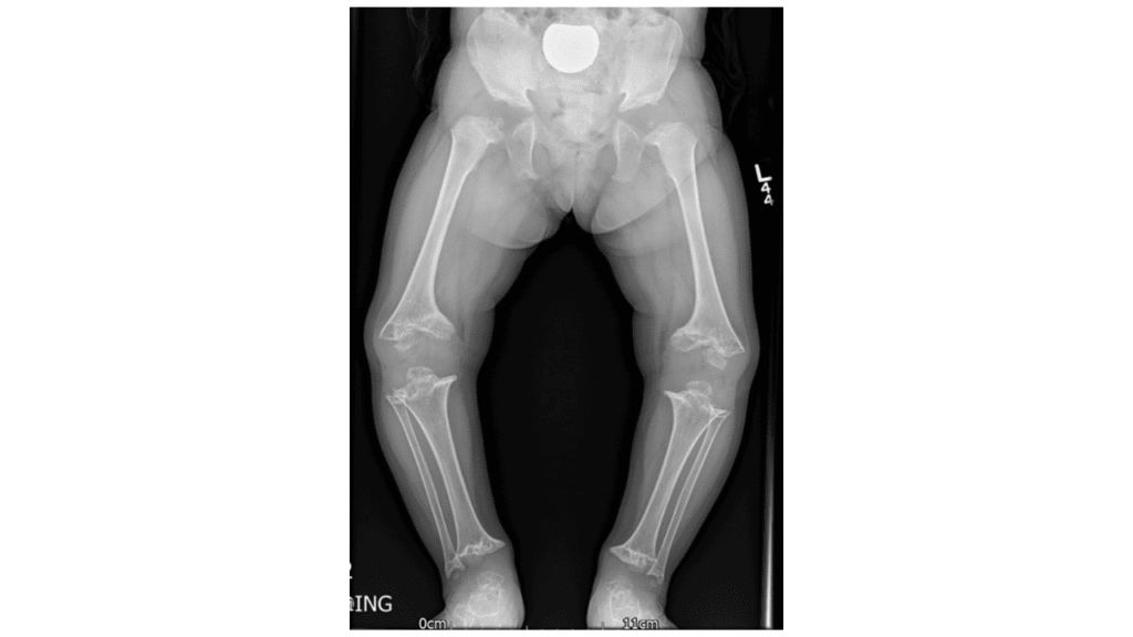
Typical radiographic findings include:
- Shortened tubular bones.
- Irregular, flattened, or underdeveloped epiphyses.
- Widened metaphyses.
- Irregular epiphyseal ossification.
- Angular deformities of the lower extremities.
- Abnormal joint alignment and congruity.
These findings reflect disturbed endochondral ossification and impaired development of the growth plates.
Spinal Abnormalities
Vertebral abnormalities may also be present. Vertebral bodies can appear oval-shaped or otherwise irregular, depending on the region of the spine and the patient’s age.
Spinal findings should be interpreted alongside the appendicular skeletal abnormalities and clinical presentation. Imaging of the spine is particularly relevant when symptoms or examination findings suggest a specific spinal complication.
Progressive Joint Deformity
As the patient grows, abnormal epiphyseal development, ligamentous laxity, and mechanical axis deviation may contribute to progressive joint deformity.
Radiographs may demonstrate irregular articular surfaces, changes in joint alignment, and degenerative changes associated with secondary osteoarthritis.
Standing alignment radiographs of the lower limbs can help quantify deformity and guide orthopedic treatment planning when indicated.
Diagnosis of Pseudoachondroplasia
Clinical Evaluation
Diagnosis begins with a detailed medical history and physical examination. Important findings include disproportionate short stature, delayed motor development, ligamentous laxity, abnormal gait, and progressive lower-limb deformity.
The clinician should assess limb proportions, joint range of motion, ligamentous stability, gait, and functional limitations. A family history of skeletal dysplasia or short stature may support the diagnosis, although many affected individuals have no known family history.
Radiographic Examination
A skeletal survey may be indicated when pseudoachondroplasia is suspected. It helps identify characteristic abnormalities of the long bones, epiphyses, metaphyses, and vertebrae.
Additional imaging should be selected according to the patient’s symptoms, examination findings, and orthopedic treatment needs.
Genetic Testing
Molecular genetic testing can confirm the diagnosis by identifying a pathogenic variant in the COMP gene.
Genetic evaluation is particularly useful when clinical or radiographic findings overlap with other skeletal dysplasias. Genetic counseling can help affected individuals and families understand inheritance, recurrence risks, and reproductive implications.
Differential Diagnosis
Pseudoachondroplasia should be distinguished from other conditions that cause disproportionate short stature and abnormal skeletal development.
Important differential diagnoses include:
- Achondroplasia: Another skeletal dysplasia associated with disproportionate short stature, but usually caused by pathogenic variants in the FGFR3 gene and characterized by a different pattern of skeletal findings.
- Multiple epiphyseal dysplasia: A group of disorders characterized by abnormal epiphyseal development, joint pain, and variable short stature; some forms are also associated with COMP variants.
- Spondyloepiphyseal dysplasia: A group of skeletal dysplasias with prominent vertebral and epiphyseal abnormalities.
- Other skeletal dysplasias: Conditions with overlapping clinical or radiographic findings that may require molecular testing for accurate classification.
Because pseudoachondroplasia and multiple epiphyseal dysplasia can both be associated with COMP, genetic results must be interpreted alongside the clinical phenotype and radiographic findings.
Orthopedic Management of Pseudoachondroplasia
Management is individualized according to the patient’s age, symptoms, degree of deformity, joint instability, and functional limitations. There is no established treatment that reverses the underlying genetic defect, so care focuses on preserving function, reducing pain, managing deformity, and addressing degenerative joint disease.
A multidisciplinary approach involving orthopedic specialists, physiotherapists, rehabilitation professionals, and clinical geneticists can help coordinate long-term care.
Conservative Management
Physical Therapy
Physical therapy is important for maintaining joint mobility, muscle strength, balance, and functional independence.
A supervised program may include:
- Range-of-motion exercises to preserve joint mobility.
- Strengthening exercises for the muscles supporting unstable joints.
- Balance and gait training.
- Functional exercises adapted to the patient’s abilities.
- Low-impact physical activities that limit excessive joint loading.
Exercise selection should be individualized. Aggressive stretching of already lax joints and activities that repeatedly provoke pain or instability should be avoided.
Pain Management
Pain management may include activity modification, appropriate analgesics, and nonsteroidal anti-inflammatory drugs when clinically indicated.
Medication selection should account for age, comorbidities, contraindications, and potential adverse effects. Persistent or worsening pain warrants assessment for progressive deformity, joint instability, or osteoarthritis rather than relying solely on medication.
Orthoses and Mobility Support
Braces, orthoses, walking aids, or other mobility supports may be considered when they improve stability, reduce symptoms, or increase walking tolerance.
Their use should be guided by an individualized assessment of limb alignment, joint instability, comfort, and functional goals.
Surgical Management
Surgery may be considered when conservative treatment does not adequately address significant deformity, instability, pain, or functional impairment.
Corrective Osteotomy
Corrective osteotomy involves cutting and realigning a bone to improve its mechanical axis and redistribute forces across a joint.
In pseudoachondroplasia, osteotomy planning can be challenging because ligamentous laxity and irregular epiphyseal surfaces may contribute to persistent instability even after bony alignment is corrected.
The appropriate correction depends on the location and magnitude of the deformity, joint congruity, skeletal maturity, and functional objectives.
In selected cases, the surgeon may need to balance anatomical alignment against the goal of providing a stable, functional limb under weight-bearing conditions. The decision requires careful individualized planning.
Management of Knee Instability
Knee instability may require a combination of strengthening, orthotic support, and surgical correction of associated deformities.
Treatment should address the underlying contributors, including ligamentous laxity, angular deformity, and joint surface abnormalities. Isolated correction of one factor may not fully resolve instability when several abnormalities coexist.
Joint Replacement
Some adults with pseudoachondroplasia develop severe secondary osteoarthritis and may eventually require joint replacement, particularly of the hips or knees.
Arthroplasty can be technically challenging because of short and sometimes narrow bones, abnormal anatomy, irregular articular surfaces, ligamentous laxity, and altered mechanical alignment.
Preoperative planning should consider implant sizing, bone stock, alignment, joint stability, and the patient’s overall functional requirements. These procedures are best planned by an orthopedic team experienced in complex joint reconstruction and skeletal dysplasia.
The decision to proceed with joint replacement depends on symptom severity, radiographic findings, functional impairment, response to conservative treatment, and the expected risks and benefits.
Prognosis and Long-Term Follow-Up
Pseudoachondroplasia is a lifelong skeletal disorder with a variable clinical course. Short stature and characteristic skeletal abnormalities persist, while limb deformity, joint instability, and degenerative joint disease may become increasingly important with age.
The rate of progression differs among individuals. Some patients maintain useful mobility for many years with conservative management, while others develop substantial pain and functional limitations requiring orthopedic intervention.
Long-term follow-up should monitor:
- Lower-limb alignment and progression of deformity.
- Joint stability and range of motion.
- Pain and signs of early osteoarthritis.
- Gait, balance, and walking endurance.
- Functional independence and participation in daily activities.
- The need for orthoses, rehabilitation, or surgical intervention.
Early recognition of deformity and individualized management may help preserve mobility and optimize quality of life.
Frequently Asked Questions About Pseudoachondroplasia
What causes pseudoachondroplasia?
Pseudoachondroplasia is usually caused by a pathogenic variant in the COMP gene on chromosome 19. Abnormal cartilage oligomeric matrix protein accumulates inside chondrocytes, disrupting cartilage function and normal bone growth.
Is pseudoachondroplasia inherited?
Yes. Pseudoachondroplasia is an autosomal dominant disorder. An affected individual has a 50% chance of passing the pathogenic variant to each child. However, some cases result from a new genetic variant without a family history.
Is pseudoachondroplasia apparent at birth?
Usually, it is not readily apparent at birth. Clinical features often become noticeable during early childhood as the child begins to grow and acquire motor milestones.
What are the main clinical features of pseudoachondroplasia?
The main features include disproportionate short stature, shortened limbs, ligamentous laxity, a waddling or wide-based gait, lower-limb malalignment, and progressive joint pain or instability.
What are the characteristic radiographic findings?
Typical findings include shortened long bones, flattened or irregular epiphyses, widened metaphyses, and variable vertebral abnormalities. Progressive malalignment and degenerative joint changes may develop with age.
How is pseudoachondroplasia diagnosed?
Diagnosis is based on clinical assessment, characteristic skeletal radiographs, and, when appropriate, molecular genetic testing for pathogenic variants in the COMP gene.
Is pseudoachondroplasia the same as achondroplasia?
No. Both conditions cause disproportionate short stature, but they are genetically and radiographically distinct. Pseudoachondroplasia is usually associated with COMP, whereas achondroplasia is usually associated with FGFR3.
Can pseudoachondroplasia cause osteoarthritis?
Yes. Abnormal joint surfaces, ligamentous laxity, and limb malalignment can increase mechanical stress on articular cartilage, contributing to early-onset secondary osteoarthritis.
What is the role of physical therapy?
Physical therapy helps maintain range of motion, strengthen muscles, improve balance, and support walking function. Programs should be individualized to avoid aggravating joint instability or pain.
Is surgery necessary for every patient?
No. Many patients are managed with monitoring, physical therapy, pain management, and supportive devices. Surgery is considered when significant deformity, instability, pain, or functional limitations warrant intervention.
Can adults with pseudoachondroplasia undergo joint replacement?
Yes. Joint replacement may be appropriate for selected adults with severe symptomatic osteoarthritis. However, abnormal bone anatomy, short stature, ligamentous laxity, and alignment problems can make the procedure more complex.
Medical References
- National Library of Medicine. COMP gene: Cartilage oligomeric matrix protein. MedlinePlus Genetics. https://medlineplus.gov/genetics/gene/comp/
- GeneReviews®. COMP-Related Pseudoachondroplasia and Multiple Epiphyseal Dysplasia. NCBI Bookshelf. https://www.ncbi.nlm.nih.gov/books/
- Briggs MD, Wright MJ. Pseudoachondroplasia. GeneReviews and related clinical genetics literature available through NCBI Bookshelf and PubMed. https://pubmed.ncbi.nlm.nih.gov/
- National Center for Biotechnology Information. Pseudoachondroplasia: clinical, radiographic, and molecular genetics literature. PubMed search. https://pubmed.ncbi.nlm.nih.gov/?term=pseudoachondroplasia+COMP
- National Center for Biotechnology Information. COMP-related skeletal dysplasia and cartilage oligomeric matrix protein. PubMed search. https://pubmed.ncbi.nlm.nih.gov/?term=COMP+related+pseudoachondroplasia+skeletal+dysplasia