Nail-patella syndrome (NPS) is a rare, inherited genetic disorder characterized by abnormalities of the fingernails, kneecaps (patellae), elbows, and pelvic bones. It is caused primarily by pathogenic variants in the LMX1B gene, which plays an essential role in the development of the limbs, kidneys, and eyes.
The condition follows an autosomal dominant inheritance pattern and affects approximately 1 in 50,000 individuals.
Although nail and skeletal abnormalities are the most recognizable features, nail-patella syndrome is a multisystem disorder. Some affected individuals develop potentially serious complications, particularly nephropathy and glaucoma.
The clinical presentation varies considerably. Some patients have mild nail abnormalities and minimal functional impairment, while others experience recurrent patellar dislocation, restricted elbow movement, chronic joint pain, or progressive kidney disease.
Early diagnosis, appropriate orthopedic management, and lifelong surveillance for renal and ophthalmological complications are essential.
This article provides a comprehensive overview of nail-patella syndrome, including its genetics, pathophysiology, clinical manifestations, radiological features, diagnosis, management, and prognosis.
What Is Nail-Patella Syndrome?
Nail-patella syndrome is a hereditary disorder affecting the normal development of several structures, particularly the nails, bones, joints, kidneys, and eyes.
It is also known as:
- Hereditary onycho-osteodysplasia (HOOD)
- Fong disease
- LMX1B-related nail-patella syndrome
The syndrome is traditionally associated with four characteristic clinical and radiological abnormalities.
The Classical Tetrad of Nail-Patella Syndrome
The four hallmark features are:
- Nail dysplasia: Hypoplastic, abnormally shaped, or absent fingernails.
- Patellar abnormalities: Small, malformed, unstable, or absent patellae.
- Elbow abnormalities: Restricted movement, radial head dysplasia, or subluxation.
- Iliac horns: Characteristic bony projections arising from the posterior iliac bones.
Not every patient demonstrates all four abnormalities.
The presence of characteristic nail changes together with patellar abnormalities should raise clinical suspicion, especially when there is a positive family history.
Bilateral iliac horns are particularly important because they are considered pathognomonic of nail-patella syndrome.
According to GeneReviews, the degree of involvement varies considerably, even among members of the same family.
Causes and Genetics of Nail-Patella Syndrome
LMX1B Gene Mutation
Nail-patella syndrome is primarily caused by a heterozygous pathogenic variant in the LMX1B gene, located on chromosome 9q34.
The LMX1B gene encodes a transcription factor involved in embryonic development.
This protein is particularly important for:
- Dorsal–ventral patterning of the limbs.
- Formation and differentiation of skeletal structures.
- Development and maintenance of kidney podocytes.
- Development of structures within the anterior segment of the eye.
Disruption of LMX1B function explains the characteristic combination of skeletal, renal, and ophthalmological abnormalities.
Most recognized disease-causing variants result in reduced or abnormal LMX1B function.
Autosomal Dominant Inheritance
Nail-patella syndrome follows an autosomal dominant inheritance pattern.
This means that a pathogenic variant affecting one copy of the LMX1B gene may be sufficient to cause the disorder.
An affected parent has a:
50% chance of transmitting the pathogenic variant to each child.
However, the severity of the disorder cannot be reliably predicted from the severity of the affected parent’s manifestations.
Some individuals inherit the condition from an affected parent, whereas others develop it because of a new, or de novo, genetic variant.
Therefore, the absence of a family history does not exclude nail-patella syndrome.
Variable Expressivity
One of the important characteristics of NPS is variable expressivity.
Affected individuals carrying the same familial pathogenic variant may demonstrate markedly different clinical manifestations.
For example, one family member may have only mild nail abnormalities, while another develops severe knee instability or nephropathy.
This variability makes clinical assessment and screening of family members particularly important.
Clinical Features of Nail-Patella Syndrome
The manifestations of nail-patella syndrome can be divided into skeletal, nail, renal, ophthalmological, and other systemic abnormalities.
1. Nail Abnormalities
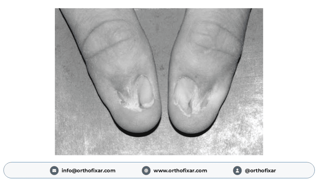
Nail abnormalities are among the earliest and most consistent features of NPS.
They occur in approximately 96-98% of affected individuals.
The fingernails are usually more severely affected than the toenails.
Common abnormalities include:
- Hypoplastic or underdeveloped nails.
- Complete absence of some nails (anonychia).
- Longitudinal ridging.
- Nail splitting or dystrophy.
- Abnormal nail pigmentation.
- Thin or irregular nail plates.
- Triangular lunulae.
Triangular Lunulae
The lunula is the pale, crescent-shaped region near the base of the nail.
In nail-patella syndrome, the lunula may appear triangular rather than semicircular.
Triangular lunulae are a highly characteristic clinical clue.
The thumbnails are usually the most severely affected.
Nail abnormalities often become progressively less pronounced from the thumb toward the little finger.
These changes may be evident from birth and provide an important opportunity for early diagnosis.
2. Patellar and Knee Abnormalities
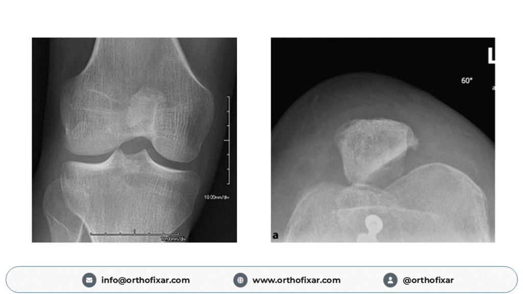
Patellar abnormalities represent one of the most clinically significant skeletal manifestations.
Knee involvement occurs in approximately 74–90% of affected individuals.
The patellae may be:
- Hypoplastic.
- Irregularly shaped.
- Abnormally positioned.
- Unstable.
- Completely absent.
Associated abnormalities of the femoral trochlea, joint alignment, and surrounding soft tissues may contribute to instability.
Clinical Presentation
Patients may experience:
- Anterior knee pain.
- Recurrent patellar subluxation.
- Patellar dislocation.
- Knee instability or giving way.
- Difficulty climbing stairs.
- Abnormal gait.
- Reduced knee function.
Some patients develop progressive patellofemoral dysfunction or early degenerative joint changes.
The severity of symptoms depends on the degree of anatomical abnormality and associated soft-tissue involvement.
Importantly, an abnormal patella does not necessarily require surgical correction if the patient remains asymptomatic.
3. Elbow Abnormalities
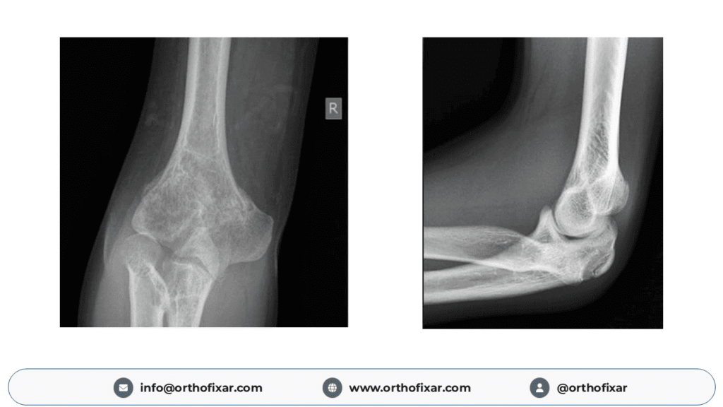
Elbow involvement occurs in approximately 70% of patients.
Common abnormalities include:
- Radial head hypoplasia or dysplasia.
- Subluxation or dislocation of the radial head.
- Limited elbow extension.
- Restricted forearm pronation and supination.
- Cubitus valgus.
- Elbow contractures.
Some patients also develop an abnormal skin fold across the anterior aspect of the elbow, known as an antecubital pterygium.
Functional Consequences
Restricted forearm rotation may interfere with daily activities requiring pronation and supination.
However, many individuals adapt well to reduced elbow mobility.
In some patients, radial head subluxation produces persistent pain or mechanical symptoms.
These cases may require specialist orthopedic evaluation.
4. Iliac Horns
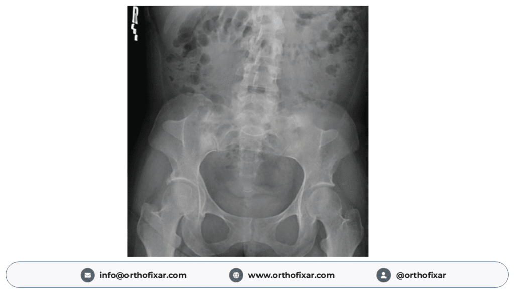
Iliac horns are one of the most distinctive radiological findings in nail-patella syndrome.
They are bilateral, cone-shaped bony projections arising from the posterior aspect of the iliac bones.
These projections typically extend posteriorly and laterally.
Iliac horns are identified on pelvic radiographs in approximately 70–80% of affected individuals, depending on the population studied.
Although their absence does not exclude NPS, their presence is considered pathognomonic.
Most iliac horns are asymptomatic and do not require treatment.
Occasionally, they may be palpable during physical examination.
Medical Education Point
The combination of:
Nail dysplasia + Patellar hypoplasia + Bilateral iliac horns
is strongly suggestive of nail-patella syndrome.
Pelvic radiographs can therefore provide valuable diagnostic information.
5. Other Skeletal Abnormalities
Additional musculoskeletal manifestations may include:
- Scoliosis.
- Increased lumbar lordosis.
- Shoulder-girdle abnormalities.
- Scapular hypoplasia.
- Foot deformities.
- Joint contractures.
- Reduced bone mineral density.
- Abnormal joint alignment.
These abnormalities vary substantially between patients.
Some individuals also experience chronic musculoskeletal pain or limitations in mobility.
Extraskeletal Manifestations of Nail-Patella Syndrome
Although the skeletal abnormalities are often the most obvious findings, the renal and ophthalmological manifestations can have greater long-term consequences.
1. Nail-Patella Syndrome Nephropathy
Kidney involvement is among the most important complications of NPS.
Approximately 30–50% of affected individuals develop some degree of renal involvement.
The earliest detectable abnormality is usually proteinuria, sometimes accompanied by hematuria.
Clinical Features of Nephropathy
Renal manifestations may include:
- Proteinuria.
- Microscopic hematuria.
- Hypertension.
- Nephrotic syndrome.
- Progressive chronic kidney disease.
- End-stage kidney disease in severe cases.
Proteinuria may develop at any age and can remain asymptomatic for many years.
Although many patients maintain satisfactory kidney function, a minority experience progressive renal impairment.
Historical studies have reported end-stage kidney disease in up to approximately 15% of affected individuals, although estimates vary considerably among cohorts.
Pathophysiology of Renal Involvement
LMX1B plays an important role in the development and maintenance of glomerular podocytes.
Abnormal LMX1B activity can affect the glomerular filtration barrier, resulting in protein leakage into the urine.
Characteristic ultrastructural abnormalities may include:
- Irregular thickening of the glomerular basement membrane.
- Abnormal collagen-like fibrils.
- Electron-lucent areas within the basement membrane.
- A characteristic moth-eaten appearance on electron microscopy.
These findings may be seen in patients with NPS-associated nephropathy.
Clinical significance: Because kidney disease may remain asymptomatic, renal screening is recommended even in patients with mild skeletal manifestations.
2. Glaucoma and Ophthalmological Abnormalities
Nail-patella syndrome is associated with an increased risk of primary open-angle glaucoma and ocular hypertension.
Glaucoma may develop at a younger age than is typical in the general population.
It is particularly important because progressive optic nerve damage can cause irreversible visual impairment.
Ophthalmological Findings
Patients may demonstrate:
- Increased intraocular pressure.
- Primary open-angle glaucoma.
- Optic nerve abnormalities.
- Visual field defects.
Glaucoma may initially be asymptomatic.
Consequently, a lack of visual complaints does not eliminate the need for ophthalmological screening.
Regular eye examinations allow earlier identification and treatment of potentially vision-threatening complications.
3. Other Systemic Manifestations
Other reported manifestations include:
- Gastrointestinal symptoms, particularly constipation.
- Reduced sensitivity to pain and temperature.
- Dental abnormalities.
- Neurological symptoms.
- Occasional seizures.
These findings are not universal and may require additional specialist assessment when clinically indicated.
Diagnosis of Nail-Patella Syndrome
The diagnosis of nail-patella syndrome is usually established through characteristic clinical findings, family history, radiological assessment, and molecular genetic testing.
Formal universal clinical diagnostic criteria have not been established.
Clinical Examination
A detailed physical examination should assess the nails, knees, elbows, spine, and other potentially affected joints.
Important findings include:
- Hypoplastic or absent nails.
- Triangular lunulae.
- Small or unstable patellae.
- Limited elbow extension.
- Reduced forearm rotation.
- Features suggesting skeletal deformity.
A three-generation family history can help identify an autosomal dominant inheritance pattern.
However, sporadic cases may occur.
Genetic Testing
Molecular genetic testing can identify a pathogenic or likely pathogenic variant in the LMX1B gene.
Testing may involve:
- Targeted LMX1B gene sequencing.
- Deletion and duplication analysis.
- A multigene panel when the diagnosis is uncertain.
- Broader genomic testing in selected cases.
Identification of a pathogenic LMX1B variant supports the diagnosis and facilitates genetic counseling.
However, a negative genetic result does not completely exclude NPS when the clinical and radiological findings are characteristic.
A variant of uncertain significance alone is not sufficient to establish the diagnosis.
Radiological Findings in Nail-Patella Syndrome
Radiological investigations are particularly valuable for evaluating skeletal abnormalities and planning orthopedic management.
1. Knee Radiographs
Plain radiographs may demonstrate:
- Hypoplastic or absent patellae.
- Patellar malposition.
- Abnormal femoral condylar morphology.
- Trochlear dysplasia.
- Associated degenerative changes.
An important consideration in young children is that normal patellar ossification centers generally become visible on radiographs between approximately three and six years of age.
Therefore, an apparently absent patella on a radiograph in a very young child should be interpreted cautiously.
2. Elbow Radiographs
Elbow radiographs may reveal:
- Dysplasia of the radial head.
- Radial head subluxation.
- Radial head dislocation.
- Abnormal joint morphology.
- Remodeling of the elbow joint.
Radiographic assessment helps explain restricted movement and mechanical symptoms.
3. Pelvic Radiographs
An anteroposterior pelvic radiograph may demonstrate the characteristic bilateral iliac horns.
These appear as bony projections extending from the iliac bones.
The identification of iliac horns strongly supports the diagnosis of nail-patella syndrome.
4. Computed Tomography (CT)
CT may be useful when detailed assessment of complex bony abnormalities is required.
It can help evaluate:
- Patellar morphology.
- Skeletal alignment.
- Complex joint anatomy.
- Bony abnormalities relevant to surgical planning.
CT is not routinely necessary for every patient and should be reserved for appropriate clinical indications.
5. Magnetic Resonance Imaging (MRI)
MRI is particularly valuable for evaluating the soft tissues and anatomical abnormalities around affected joints.
It can demonstrate:
- Patellofemoral malalignment.
- Trochlear dysplasia.
- Cartilage abnormalities.
- Meniscal abnormalities.
- Ligament and soft-tissue pathology.
- Abnormal fibrous or synovial structures within the knee.
MRI is especially useful before complex reconstructive surgery.
Detailed preoperative imaging helps orthopedic surgeons understand the anatomical abnormalities and select appropriate surgical procedures.
Differential Diagnosis
Several other conditions may produce abnormal nails, absent patellae, or skeletal abnormalities.
The differential diagnosis may include:
- Isolated congenital patellar aplasia or hypoplasia.
- Small patella syndrome.
- Meier–Gorlin syndrome.
- Genitopatellar syndrome.
- Other inherited skeletal dysplasias.
- Disorders associated with congenital nail abnormalities.
The combination of characteristic nail changes, patellar abnormalities, elbow involvement, and iliac horns helps distinguish NPS from other conditions.
Genetic evaluation may be needed when the clinical presentation is atypical.
Treatment and Management of Nail-Patella Syndrome
There is currently no curative treatment for nail-patella syndrome.
Management is individualized and focuses on preserving joint function, treating symptoms, preventing complications, and monitoring potentially affected organ systems.
A multidisciplinary approach is often appropriate.
1. Conservative Orthopedic Management
Conservative treatment is generally the initial approach for patients with mild or moderate musculoskeletal symptoms.
Management may include:
- Physiotherapy.
- Muscle-strengthening exercises.
- Joint stabilization exercises.
- Activity modification.
- Appropriate analgesia.
- Splinting or bracing when indicated.
Physiotherapy
Physiotherapy aims to improve function, optimize muscle strength, and reduce pain.
For patients with knee instability, rehabilitation may focus on quadriceps strengthening, hip muscle conditioning, and functional stabilization.
However, exercise programs must be adapted to the patient’s anatomical abnormalities.
Patients with significant joint deformities may require imaging and specialist orthopedic assessment before intensive rehabilitation.
Pain Management
Pain relief should be individualized.
Nonsteroidal anti-inflammatory drugs (NSAIDs) require particular caution.
Long-term NSAID use should generally be avoided in patients with NPS because of the potential adverse effects on kidney function.
Analgesic selection should account for the patient’s renal status and other medical conditions.
2. Surgical Treatment of Patellar Instability
Surgery may be considered in patients with:
- Recurrent symptomatic patellar dislocation.
- Significant patellar instability.
- Persistent anterior knee pain despite appropriate conservative treatment.
- Progressive functional impairment.
- Structural abnormalities requiring correction.
Patellar stabilization can be challenging because of the combination of abnormal bony anatomy, malalignment, and soft-tissue abnormalities.
Depending on the individual presentation, surgical options may include:
- Soft-tissue realignment.
- Patellar stabilization procedures.
- Medial patellofemoral ligament reconstruction.
- Correction of selected associated deformities.
- Other reconstructive procedures tailored to the patient’s anatomy.
The selection of a procedure requires careful assessment of the patella, femoral trochlea, surrounding ligaments, and overall limb alignment.
Not every patient with patellar hypoplasia requires surgery.
The decision should be based primarily on symptoms, instability, functional impairment, and specialist assessment.
3. Surgical Treatment of Elbow Abnormalities
Elbow surgery is less commonly required.
Many patients tolerate restricted forearm rotation without major functional disability.
Surgical treatment may be considered when there is severe pain, significant mechanical dysfunction, or disabling deformity.
In carefully selected cases, procedures such as soft-tissue release or radial head excision have been reported.
However, radial head excision is not a routine treatment for NPS.
Its potential benefits and risks must be carefully assessed by an experienced orthopedic surgeon.
The published evidence for orthopedic surgery in NPS is limited, consisting largely of case reports and small patient series. Therefore, treatment decisions must remain individualized.
4. Management of Nephropathy
Patients with renal abnormalities should be assessed by a nephrologist.
Treatment depends on the severity and progression of renal disease.
Management may include:
- Blood pressure control.
- Monitoring urinary protein excretion.
- Evaluation of kidney function.
- Treatment of proteinuria.
- Management of chronic kidney disease.
Angiotensin-converting enzyme (ACE) inhibitors may help control hypertension and reduce proteinuria in appropriate patients.
Treatment requires medical supervision, particularly in children and during pregnancy.
Patients who develop advanced kidney failure may require dialysis or kidney transplantation.
5. Management of Glaucoma
Glaucoma management should be supervised by an ophthalmologist.
Treatment may include:
- Intraocular pressure-lowering medications.
- Laser procedures.
- Glaucoma surgery when indicated.
Early detection is essential because glaucoma-related optic nerve damage is generally irreversible.
Long-Term Monitoring and Follow-Up
Because NPS is a multisystem disorder, regular medical surveillance is necessary even when musculoskeletal symptoms are mild.
The updated GeneReviews recommendations emphasize annual screening for several important complications.
Recommended Follow-Up Assessments
| System | Recommended Evaluation | Frequency |
|---|---|---|
| Musculoskeletal | Assessment of joint function, gait, mobility, and spinal abnormalities | At least annually |
| Renal | Urinalysis and first-morning urine albumin-to-creatinine ratio | At least annually |
| Cardiovascular | Blood pressure measurement | At least annually |
| Ophthalmological | Screening for glaucoma | At least annually once the child can cooperate |
| Dental | Dental examination | Every 6 months |
| Neurological | Assessment for relevant symptoms or sensory abnormalities | At least annually |
| Bone health | Bone mineral density evaluation when indicated | Based on clinical findings |
Patients with abnormal kidney screening results should be referred for nephrology assessment.
An ophthalmologist should determine whether more frequent eye examinations are needed.
Surveillance should be individualized according to clinical findings and previous test results.
Important: Normal kidney function and absence of glaucoma at the time of diagnosis do not eliminate the need for future screening.
Complications of Nail-Patella Syndrome
Potential complications include:
Musculoskeletal Complications
- Recurrent patellar dislocation.
- Chronic anterior knee pain.
- Reduced joint mobility.
- Elbow dysfunction.
- Degenerative joint disease.
- Functional limitations.
Renal Complications
- Persistent proteinuria.
- Hypertension.
- Chronic kidney disease.
- Nephrotic syndrome.
- Kidney failure in severe cases.
Ophthalmological Complications
- Ocular hypertension.
- Progressive glaucoma.
- Optic nerve damage.
- Permanent visual impairment.
The probability and severity of complications vary substantially between affected individuals.
Early recognition and appropriate follow-up can help reduce preventable morbidity.
Prognosis of Nail-Patella Syndrome
The long-term outlook depends mainly on the severity of associated complications.
Many individuals with mild skeletal abnormalities and preserved kidney and eye health can maintain a good quality of life.
Some patients experience chronic joint symptoms or limitations in mobility.
Renal involvement represents an important determinant of long-term health because progressive nephropathy can occasionally lead to kidney failure.
Similarly, untreated glaucoma can result in permanent visual impairment.
Therefore, the prognosis is influenced not only by musculoskeletal disease but also by early detection and management of renal and ophthalmological complications.
The condition demonstrates marked variability, and the clinical course cannot be predicted reliably from the appearance of the nails or severity of patellar abnormalities alone.
Nail-Patella Syndrome: Important Medical Examination Points
For medical students and healthcare professionals, the following points are particularly important:
- Inheritance: Autosomal dominant.
- Causative gene: LMX1B on chromosome 9q34.
- Classical tetrad: Nail dysplasia, patellar abnormalities, elbow abnormalities, and iliac horns.
- Characteristic nail finding: Triangular lunulae.
- Characteristic radiological finding: Bilateral iliac horns.
- Common knee manifestation: Patellar hypoplasia with instability or dislocation.
- Elbow manifestation: Restricted pronation and supination, sometimes associated with radial head dysplasia or subluxation.
- Important renal complication: Proteinuria and progressive nephropathy.
- Important ophthalmological complication: Primary open-angle glaucoma.
- Management: Symptom-directed orthopedic treatment with ongoing kidney and eye surveillance.
High-Yield Clinical Pearl
A patient presenting with hypoplastic fingernails, recurrent patellar instability, and bilateral iliac horns on pelvic radiography should be evaluated for nail-patella syndrome.
The diagnosis should prompt assessment for associated renal disease and glaucoma, even in the absence of symptoms.
Frequently Asked Questions About Nail-Patella Syndrome
Is nail-patella syndrome hereditary?
Yes. Nail-patella syndrome is usually inherited in an autosomal dominant manner.
An affected individual has a 50% chance of passing the causative genetic variant to each child.
However, new genetic variants can also cause the disorder in individuals without an affected parent.
Which gene causes nail-patella syndrome?
The principal gene associated with nail-patella syndrome is LMX1B, located on chromosome 9q34.
This gene encodes a transcription factor involved in limb, kidney, and eye development.
What is the most characteristic radiological finding in nail-patella syndrome?
The most characteristic radiological finding is the presence of bilateral iliac horns.
These are bony projections extending from the posterior iliac bones and are considered pathognomonic for the syndrome.
Are iliac horns present in every patient?
No. Iliac horns are present in approximately 70–80% of affected individuals, depending on the study.
Their absence does not exclude the diagnosis.
Can nail-patella syndrome affect the kidneys?
Yes. Approximately 30–50% of affected individuals develop some degree of renal involvement.
Proteinuria is usually the earliest finding, although some patients develop progressive chronic kidney disease.
Does everyone with nail-patella syndrome develop glaucoma?
No. Glaucoma does not occur in every affected individual.
However, patients with NPS have an increased risk of glaucoma and ocular hypertension, making regular ophthalmological screening important.
Can nail-patella syndrome be cured?
There is currently no cure for the underlying genetic condition.
Treatment focuses on controlling symptoms, maintaining joint function, and identifying or managing complications involving the kidneys, eyes, and skeletal system.
Is surgery necessary for nail-patella syndrome?
Not always.
Many patients can be managed conservatively with physiotherapy, bracing, activity modification, and appropriate pain management.
Surgical treatment is generally reserved for selected patients with significant joint instability, persistent symptoms, or functional impairment.
What is the life expectancy of someone with nail-patella syndrome?
There is no single life-expectancy estimate that applies to everyone with NPS.
Many affected individuals have mild disease, while some develop serious renal complications.
Long-term outcomes depend largely on individual manifestations, particularly the presence and progression of kidney disease.
Conclusion
Nail-patella syndrome is a rare autosomal dominant genetic disorder caused primarily by pathogenic variants in the LMX1B gene.
It is characterized by a classical combination of nail dysplasia, patellar abnormalities, elbow deformities, and iliac horns.
The presence of bilateral iliac horns on pelvic radiographs is a particularly important diagnostic finding.
Although orthopedic abnormalities are the most recognizable manifestations, clinicians must also consider extraskeletal complications, especially nephropathy and glaucoma.
Diagnosis involves clinical examination, family history, characteristic radiological findings, and molecular genetic testing when appropriate.
Management is individualized and typically begins with conservative orthopedic treatment. Surgical correction may be required in selected patients with significant instability or functional impairment.
Regular renal and ophthalmological surveillance remains an essential component of long-term care.
Recognition of the characteristic clinical features enables earlier diagnosis, appropriate multidisciplinary treatment, and better prevention of complications.
References and Trusted Medical Sources
The following sources are indexed in PubMed or hosted by the National Center for Biotechnology Information (NCBI).
- 1. Hoover-Fong JE, McIntosh I. Nail-Patella Syndrome. GeneReviews®. NCBI Bookshelf. Updated June 2, 2026. A comprehensive resource covering molecular genetics, clinical manifestations, diagnostic evaluation, management, genetic counseling, and surveillance. Read GeneReviews on NCBI Bookshelf
- 2. Beaugé A, Sbampato V, Vazquez O, et al. Orthopedic manifestations and management of nail-patella syndrome: a narrative review. Frontiers in Pediatrics. 2026;14:1744875. DOI: 10.3389/fped.2026.1744875 PMID: 41716707. A recent review covering orthopedic findings, radiological investigations, surgical management, and treatment outcomes. Read the 2026 Review on PubMed
- 3. Sweeney E, Fryer A, Mountford R, Green A, McIntosh I. Nail patella syndrome: a review of the phenotype aided by developmental biology. Journal of Medical Genetics. 2003;40(3):153–162. DOI: 10.1136/jmg.40.3.153 PMID: 12624132. An important clinical review describing phenotypic variability and systemic manifestations of NPS. Read the Study on PubMed
- 4. Lovelace PD, May LA. Nail-Patella Syndrome. StatPearls. NCBI Bookshelf. A clinical reference describing the genetics, pathophysiology, diagnostic evaluation, imaging findings, and management of nail–patella syndrome. Read StatPearls on NCBI Bookshelf
- 5. Galloway G, Vivian A. An ophthalmic screening protocol for nail-patella syndrome. Journal of Pediatric Ophthalmology and Strabismus. 2003;40(1):51–53. PMID: 12580274. A publication addressing the importance of ophthalmological assessment in nail–patella syndrome. Read the Publication on PubMed