Rickets is a metabolic bone disorder affecting growing children and adolescents, characterized by defective mineralization of the growth plate and newly formed bone. The disease results in skeletal deformities, impaired growth, bone pain, and an increased risk of fractures. Although nutritional vitamin D deficiency remains the most common cause worldwide, inherited disorders such as XLH rickets (X-linked hypophosphatemia) and chronic kidney disease are increasingly recognized causes.
Understanding the rickets definition, identifying rickets symptoms, recognizing different rickets types, and initiating appropriate rickets treatment are essential to prevent permanent skeletal deformities and optimize long-term outcomes.
What is Rickets?
The rickets definition is:
Rickets is a disorder of defective mineralization of the growth plate cartilage and osteoid in growing children, leading to bone deformities, impaired growth, and skeletal weakness.
Unlike osteomalacia, which affects adults after growth plate closure, rickets occurs only in children with open epiphyseal growth plates.
The condition primarily results from disturbances in calcium, phosphate, or vitamin D metabolism.
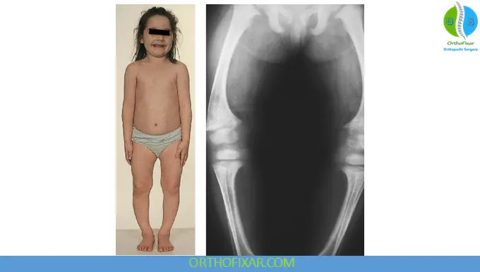
Normal Bone Mineralization
Normal bone growth requires adequate amounts of:
- Calcium
- Phosphate
- Vitamin D
- Healthy growth plates
- Normal kidney function
- Appropriate parathyroid hormone (PTH) regulation
Vitamin D increases intestinal absorption of calcium and phosphate, while phosphate is essential for hydroxyapatite crystal formation.
Failure of these mechanisms leads to poorly mineralized bone and widened growth plates.
See Also: Bone Formation & Development
Rickets Causes
The rickets causes vary depending on the underlying metabolic abnormality.
Nutritional Vitamin D Deficiency
The most common cause worldwide.
Risk factors include:
- Limited sunlight exposure
- Exclusive breastfeeding without supplementation
- Dark skin pigmentation
- Poor dietary intake
- Malabsorption disorders
H3: Calcium Deficiency
May occur despite normal vitamin D levels.
Common in regions with diets low in dairy products.
Phosphate Deficiency
Can result from:
- Renal phosphate wasting
- Inherited disorders
- Fanconi syndrome
Genetic Disorders
Inherited causes include:
- XLH rickets
- Vitamin D–dependent rickets type I
- Vitamin D–dependent rickets type II
- Hereditary hypophosphatemic disorders
Chronic Kidney Disease
Reduced vitamin D activation and impaired phosphate regulation contribute to renal rickets.
Malabsorption Disorders
Examples include:
- Celiac disease
- Inflammatory bowel disease
- Cystic fibrosis
- Short bowel syndrome
H2: Risk Factors
Children at increased risk include:
- Premature infants
- Exclusively breastfed infants without vitamin D supplementation
- Limited outdoor activity
- Darker skin pigmentation
- Poor nutrition
- Chronic liver disease
- Chronic kidney disease
- Family history of inherited rickets
Rickets Types
Several rickets types are recognized.
Nutritional Rickets
The most common form.
Caused by vitamin D and/or calcium deficiency.
Hypophosphatemic Rickets
Characterized by renal phosphate wasting.
Includes:
- XLH rickets
- Autosomal dominant hypophosphatemic rickets
- Autosomal recessive hypophosphatemic rickets
Vitamin D–Dependent Rickets
Caused by inherited defects in vitamin D metabolism or receptor function.
Includes:
- Type I
- Type II
Renal Rickets
Occurs secondary to chronic kidney disease.
Rickets Symptoms
The severity of rickets symptoms depends on age, duration, and underlying cause.
Common symptoms include:
Skeletal Symptoms
- Bone pain
- Delayed walking
- Waddling gait
- Progressive limb deformities
- Bowed legs (genu varum)
- Knock knees (genu valgum)
Growth Abnormalities
- Short stature
- Delayed growth
- Delayed motor milestones
Skull Changes
- Frontal bossing
- Delayed fontanelle closure
- Craniotabes in infants
Chest Deformities
- Rachitic rosary
- Harrison sulcus
- Pectus deformities
Limb Deformities
- Wrist widening
- Ankle widening
- Long bone bowing
Muscle Symptoms
- Muscle weakness
- Fatigue
- Difficulty climbing stairs
Hypocalcemia Symptoms
Severe deficiency may cause:
- Tetany
- Seizures
- Laryngospasm
- Cardiac arrhythmias
Physical Examination
Typical findings include:
- Bowed lower limbs
- Wide wrists
- Enlarged costochondral junctions
- Delayed dentition
- Poor muscle tone
- Gait abnormalities
- Skeletal tenderness
- Growth retardation
Diagnosis of Rickets
Diagnosis combines clinical findings, laboratory investigations, and imaging.
Laboratory Tests
Typical investigations include:
- Serum calcium
- Serum phosphate
- Alkaline phosphatase (usually elevated)
- Parathyroid hormone
- 25-hydroxyvitamin D
- 1,25-dihydroxyvitamin D (selected cases)
- Renal function
- Urinary phosphate
Radiographic Findings
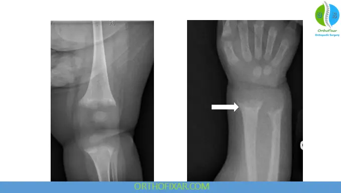
Classic X-ray findings include:
- Growth plate widening
- Metaphyseal cupping
- Metaphyseal fraying
- Metaphyseal splaying
- Delayed mineralization
- Bowing deformities
Common imaging sites:
- Wrist
- Knee
Genetic Testing
Recommended for suspected inherited disorders including XLH rickets.
Differential Diagnosis
Conditions that may resemble rickets include:
- Osteomalacia
- Osteogenesis imperfecta
- Skeletal dysplasia
- Blount disease
- Physiologic bowing
- Renal osteodystrophy
Rickets Treatment
The appropriate rickets treatment depends on the underlying cause.
Nutritional Rickets
Management includes:
- Vitamin D supplementation
- Oral calcium replacement
- Nutritional counseling
- Adequate sunlight exposure
Most children show radiographic improvement within several months.
Treatment of Hypophosphatemic Rickets
Treatment typically includes:
- Oral phosphate supplements
- Active vitamin D analogs (calcitriol or alfacalcidol)
Newer targeted therapy:
- Burosumab, a monoclonal antibody against fibroblast growth factor 23 (FGF23), is approved for many patients with XLH rickets and has demonstrated significant improvements in phosphate homeostasis, growth, and skeletal outcomes.
XLH Rickets Treatment
Current management includes:
- Burosumab
- Oral phosphate (selected patients)
- Active vitamin D analogs
- Orthopedic correction when necessary
- Regular follow-up with pediatric endocrinology
Orthopedic Management
Orthopedic intervention may be required for:
- Severe deformity
- Progressive bowing
- Limb length discrepancy
- Persistent deformity despite medical therapy
Treatment options include:
- Guided growth
- Corrective osteotomy
- Physical therapy
Complications
Untreated rickets disease may lead to:
- Permanent limb deformity
- Short stature
- Recurrent fractures
- Chronic pain
- Dental abnormalities
- Delayed motor development
- Pelvic deformities
- Reduced quality of life
Prevention
Preventive measures include:
- Vitamin D supplementation during infancy
- Adequate dietary calcium
- Safe sunlight exposure
- Maternal vitamin D optimization during pregnancy
- Early screening in high-risk children
Prognosis
The prognosis depends on the underlying etiology and timing of treatment.
- Nutritional rickets generally has an excellent prognosis when treated early.
- Genetic forms require lifelong follow-up.
- Early diagnosis significantly reduces permanent skeletal deformities.
Frequently Asked Questions
What is the difference between rickets and osteomalacia?
Rickets occurs in children before growth plate closure, whereas osteomalacia affects adults after skeletal maturity.
What causes rickets?
The most common causes are vitamin D deficiency, calcium deficiency, phosphate disorders, inherited diseases such as XLH rickets, and chronic kidney disease.
Is rickets curable?
Most nutritional cases are highly treatable with vitamin D and calcium. Genetic forms require long-term management.
What are the earliest rickets symptoms?
Early symptoms include delayed walking, bone pain, widened wrists, muscle weakness, and growth delay.
Can adults develop rickets?
No. Adults develop osteomalacia, not rickets.
Key Points
- Rickets disease is a disorder of defective bone mineralization in growing children.
- Vitamin D deficiency remains the leading global cause.
- Rickets symptoms include bone pain, bowed legs, widened wrists, and delayed growth.
- Rickets causes include nutritional deficiency, genetic disorders, renal disease, and phosphate wasting.
- Rickets treatment depends on the underlying etiology and may involve vitamin D, calcium, phosphate supplementation, or targeted therapy.
- XLH rickets is the most common inherited form and is caused by mutations affecting phosphate regulation.
- Early diagnosis prevents permanent skeletal deformities and improves long-term outcomes.
References & More
- Munns CF, Shaw N, Kiely M, et al. Global Consensus Recommendations on Prevention and Management of Nutritional Rickets. Journal of Clinical Endocrinology & Metabolism. 2016. Link
- Carpenter TO, Whyte MP, Imel EA, et al. Burosumab Therapy in Children with X-Linked Hypophosphatemia. New England Journal of Medicine. 2018. Link
- National Center for Biotechnology Information (NCBI). Vitamin D and Bone Health.
- National Institutes of Health Office of Dietary Supplements. Vitamin D Fact Sheet for Health Professionals.
- MedlinePlus. Rickets.
- GeneReviews. X-Linked Hypophosphatemia (XLH).
- UpToDate. Nutritional rickets and vitamin D deficiency in children.