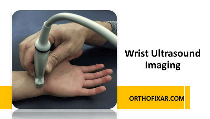
Wrist Ultrasound Imaging has become an invaluable diagnostic tool in modern medicine, offering real-time, dynamic visualization of complex anatomical structures. This imaging modality provides excellent soft tissue contrast and allows for immediate assessment of tendons, nerves, ligaments, and vascular structures without radiation exposure. The wrist’s superficial anatomy makes it particularly well-suited for ultrasound evaluation, enabling clinicians to diagnose a wide range of pathologies from carpal tunnel syndrome to tendon injuries.
Anterior Wrist Ultrasound Imaging
Carpal Tunnel Assessment
The carpal tunnel represents one of the most clinically significant areas for ultrasound evaluation on the anterior wrist. This fibro-osseous tunnel serves as a conduit for the median nerve and nine flexor tendons as they pass from the forearm into the hand.
Key Anatomical Landmarks:
- Proximal carpal tunnel: The scaphoid (laterally) and pisiform bone (medially) serve as essential bony landmarks
- Distal landmarks: The trapezium and hook of the hamate define the distal boundary
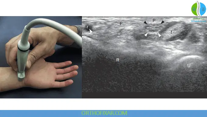
Scanning Technique: Begin by placing the transducer along the proximal carpal tunnel in the short axis. This positioning allows visualization of multiple structures simultaneously, including the median nerve, flexor carpi radialis, flexor digitorum superficialis, and the tendons of the flexor digitorum profundus. The radius and ulna provide additional bony landmarks for orientation.
Dynamic Wrist Ultrasound evaluation enhances diagnostic accuracy by having the patient perform finger flexion and extension movements. This technique reveals tendon gliding patterns and can identify adhesions or mechanical restrictions within the carpal tunnel.
See Also: Wrist Anatomy: Bones, Ligaments & Joints
Median Nerve Evaluation
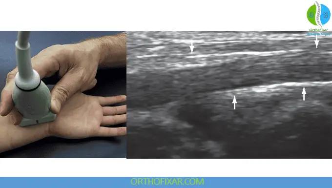
The median nerve appears as a “honeycomb” pattern when viewed in short axis, displaying hypoechoic fascicles surrounded by hyperechoic epineurial connective tissue. This appearance helps differentiate the nerve from surrounding tendinous structures.
Morphological Changes:
- Proximal: Oval-shaped configuration
- Distal: Flattened appearance at the level of the hook of the hamate
Pathological Assessment: Normal median nerve cross-sectional area at the wrist measures approximately 9 mm², increasing to 14 mm² in patients with carpal tunnel syndrome. Measurements should be standardized at the level of the distal radius or pisiform to ensure reliability and reproducibility.
Long Axis Visualization: Rotating the transducer 90° provides long axis views where nerve fascicles are clearly delineated. This orientation facilitates identification of nerve compression, characterized by:
- Enlargement of nerve diameter at the compression site
- Proximal nerve swelling
- Echotexture changes indicating pathology
Flexor Tendon Complex
The flexor tendons begin their course at the wrist crease and can be followed both proximally and distally. The superficialis and profundus muscles surround the median nerve at the wrist level.
Synovial Sheath Characteristics: Flexor digitorum tendons are enveloped by synovial sheaths that appear sonographically as thin, echogenic, fluid-filled structures. These sheaths facilitate smooth tendon gliding and can become inflamed in pathological conditions.
Anatomical Variations:
- Palmaris longus: Runs superficial to the carpal tunnel near the wrist midline
- Flexor carpi ulnaris: Located superficial and on the ulnar side of the wrist
Radial Aspect Assessment
The radial side of the anterior wrist provides access to several important structures. With the transducer positioned in long axis lateral to the median nerve, the flexor carpi radialis can be observed attaching to the second and third metacarpals. In this plane, the tendon exhibits a hypoechoic appearance with a characteristic fibrillar pattern.
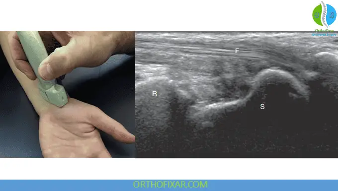
Bony Landmark Identification: The distal radius and scaphoid appear as hyperechoic bone contours. Clear visualization of the scaphoid surface allows detection of irregularities that may indicate fracture, making ultrasound a valuable tool for acute trauma assessment.
Vascular Structures: In short axis orientation, the radial artery and vein can be identified just radial to the flexor carpi radialis tendon, providing important information about vascular integrity.
Ulnar Aspect Evaluation
The ulnar side of the anterior wrist houses the ulnar nerve, artery, and vein. The pisiform bone serves as a crucial landmark for locating these structures. The ulnar nerve appears between the pisiform and ulnar artery, displaying the same honeycomb pattern of hypoechoic fascicles surrounded by hyperechoic connective tissue seen in the median nerve.
Clinical Significance of the Hook of Hamate: Moving distally, the hook of the hamate becomes visible and represents a critical anatomical region. Hamate fractures commonly occur during:
- Falls on outstretched hands
- Golf swings
- Baseball batting
These injuries can cause compression of surrounding nerves and arteries, making ultrasound assessment valuable for both diagnosis and monitoring.
Ulnar Nerve Branches: In short axis, the ulnar nerve branches can be observed coursing around the hamate, with the deep motor branch on the ulnar side and the sensory branch superficial to the hamate.
See Also: Wrist Pain Causes
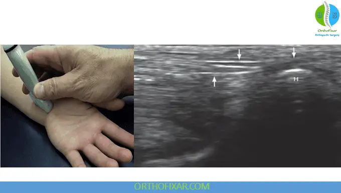
Posterior (Dorsal) Wrist Ultrasound Imaging
Extensor Tendon Compartments
The dorsal wrist contains six extensor compartments, each of them contains specific tendons that can be systematically evaluated with ultrasound. Lister’s tubercle serves as the primary landmark, appearing as a hyperechoic bony prominence near the midline of the dorsal wrist.
See Also: Extensor Compartments of the Wrist
Systematic Scanning Approach: Starting at Lister’s tubercle and moving ulnarly, the following structures can be identified:
- Extensor pollicis longus
- Extensor indicis
- Multiple tendons of the extensor digitorum
- Extensor digiti minimi
- Extensor carpi ulnaris (most ulnar)
Moving radially from Lister’s tubercle reveals:
Dynamic Assessment Benefits: Long axis visualization combined with dynamic movement improves detection of:
- Tendon end retraction following rupture
- Tendon subluxation or dislocation
- Impingement syndromes
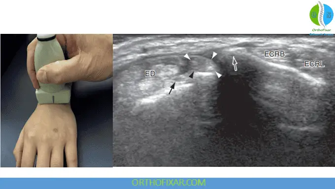
Pathological Findings
Tendinosis Characteristics:
- Variable areas of decreased echogenicity
- Tendon thickening
- Associated sheath effusion
Tendon Tears:
- Complete or partial discontinuity of tendon fibers
- Fluid interposition at the tear site
- Retraction of tendon ends in complete ruptures
Triangular Fibrocartilage Complex (TFCC)
Located on the ulnar side of the dorsal wrist, the TFCC sits between the carpal bones and the ulna. This structure is crucial for load transmission and wrist stability, making its assessment important in patients with ulnar-sided wrist pain.
See Also: Triangular Fibrocartilage Complex Injury
Specialized Thumb Ligament Assessment
Ulnar Collateral Ligament (UCL) – “Gamekeeper’s Thumb”
UCL injuries of the first metacarpophalangeal joint result from forceful hyperabduction of the thumb, commonly occurring in:
- Contact sports
- Skiing accidents (ski pole mechanism)
- Falls
Ultrasound Appearance: Injured ligaments appear hyperechoic and swollen compared to normal tissue. A smaller “hockey stick” transducer is optimal for this assessment due to the confined anatomical space.
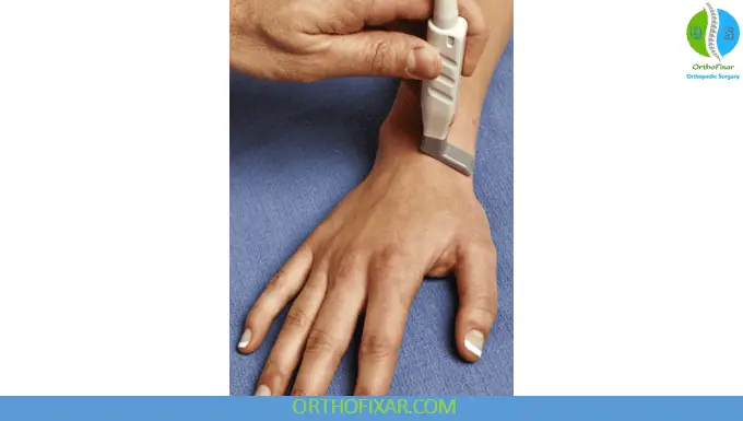
Stener Lesion Recognition: This injury occurs when a full-thickness UCL tear results in retraction and displacement of the torn end under the adductor aponeurosis. The characteristic “yo-yo on a string” appearance describes:
- “Yo-yo”: The balled-up, proximally retracted UCL
- “String”: The adductor aponeurosis
Dynamic evaluation in long axis provides superior visualization of both the ligament and adductor aponeurosis, enabling accurate diagnosis of this injury.
Radial Collateral Ligament Assessment
Although less common than UCL injuries, radial collateral ligament tears require significant force and result in joint instability with progressive deterioration. The ultrasound appearance mirrors that of UCL injuries, but the clinical implications may be more severe due to the biomechanical importance of the radial stabilizing structures.
Clinical Applications and Diagnostic Value
Wrist ultrasound imaging provides numerous advantages in clinical practice:
Immediate Assessment: Real-time wrist ultrasound imaging allows for immediate correlation between symptoms and anatomical findings.
Dynamic Evaluation: Movement during scanning reveals functional abnormalities not apparent on static imaging.
Cost-Effective: Compared to MRI, wrist ultrasound offers an economical alternative for many wrist pathologies.
Patient Comfort: Non-invasive imaging without ionizing radiation or contrast agents.
Guided Interventions: Real-time visualization enables precise needle placement for injections or aspirations.
Conclusion
Ultrasound imaging of the wrist has evolved into an essential diagnostic tool that complements clinical examination and provides detailed anatomical information. Mastery of wrist ultrasound techniques enables clinicians to diagnose a wide range of pathologies efficiently and accurately. The combination of systematic scanning protocols, understanding of normal anatomical appearances, and recognition of pathological changes makes wrist ultrasound an invaluable skill in modern medical practice.
As technology continues to advance and transducer resolution improves, the diagnostic capabilities of wrist ultrasound will likely expand further, potentially replacing more expensive imaging modalities for many clinical scenarios. Healthcare providers who develop expertise in wrist ultrasound imaging will be better equipped to provide timely, accurate diagnoses and improve patient outcomes in musculoskeletal medicine.
References & More
- Olchowy C, Solinski D, Lasecki M, et al. Wrist ultrasound examination – scanning technique and ultrasound anatomy. Part 2: ventral wrist. J Ultrason. 2017;17(69):123–128. PubMed
- Starr HM, Sedgley MD, Means KR, Murphy MS. Ultrasonography for hand and wrist conditions. J Am Acad Orthop Surg. 2016;24(8):544–554. PubMed
- Wiesler ER, Chloros GD, Cartwright MS, et al. The use of diagnostic ultrasound in carpal tunnel syndrome. J Hand Surg Am. 2006;31(5):726–732. PubMed
- Sofka CM. Ultrasound of the hand and wrist. Ultrasound Q. 2014;30(3):184–192. PubMed
- Chen P, Maklad N, Radwine M, et al. Dynamic high resolution sonograph of the carpal tunnel. Am J Roentgenol. 1997;168(2):533–537. PubMed
- Chiavaras MM, Jacobson JA, Yablon CM, et al. Pitfalls in wrist and hand ultrasound. Am J Roentgenol. 2014;203(3):531–540. PubMed
- Daenen B, Houben G, Bauduin E, et al. Sonography in wrist tendon pathology. J Clin Ultrasound. 2004;32(9):462–469. PubMed
- Jones MH, England SJ, Muwanga CL, Hildreth T. The use of ultrasound in the diagnosis of injuries of the ulnar collateral ligament of the thumb. J Hand Surg Br. 2000;25(1):29–32. PubMed
- Melville DM, Jacobson JA, Fessell DP. Ultrasound of the thumb ulnar collateral ligament: techniques and pathology. Am J Roentgenol. 2014;202(2):W168. PubMed
- Ebrahim FS, De Maeseneer M, Jager T, et al. Ultrasound diagnosis of UCL tears of the thumb and Stener lesions: technique, pattern-based approach, and differential diagnosis. Radiographics. 2006;26:1007–1020. PubMed
