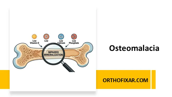
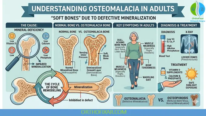
Osteomalacia is a metabolic bone disorder characterized by defective mineralization of mature bone matrix (osteoid) after the closure of growth plates. The condition results in soft, weak bones that are more susceptible to pain, deformities, and fractures.
Although vitamin D deficiency remains the most common cause of osteomalacia worldwide, several other disorders affecting calcium, phosphate, or vitamin D metabolism may also contribute to its development. Osteomalacia is often underdiagnosed because its symptoms can be subtle and nonspecific during the early stages.
This article reviews the pathophysiology, causes, clinical manifestations, diagnosis, treatment, and prevention of osteomalacia based on current medical evidence.
What is Osteomalacia?
Osteomalacia refers to inadequate mineralization of the bone matrix in adults. In children, a similar mineralization defect affects the growth plates and is known as rickets.
Normal bone mineralization requires adequate levels of:
- Vitamin D
- Calcium
- Phosphate
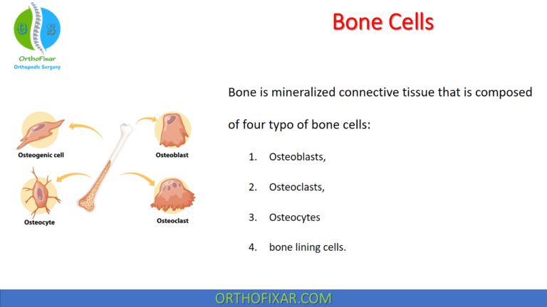
- Functional osteoblast activity
When these factors are disrupted, newly formed osteoid fails to mineralize properly, resulting in soft and fragile bones.
See Also: Bone Formation
Epidemiology
Osteomalacia remains a significant global health concern despite advances in nutritional awareness.
Individuals at increased risk include:
- Elderly adults
- Institutionalized patients
- Individuals with limited sunlight exposure
- Patients with malabsorption syndromes
- Chronic kidney disease patients
- Chronic liver disease patients
- Individuals taking anticonvulsant medications
- People with poor dietary vitamin D intake
Vitamin D deficiency remains the leading cause of osteomalacia worldwide.
Pathophysiology of Osteomalacia
Bone strength depends on proper mineralization of the collagen-rich osteoid matrix.
In osteomalacia:
- Vitamin D deficiency reduces intestinal calcium absorption.
- Hypocalcemia stimulates parathyroid hormone (PTH) secretion.
- Secondary hyperparathyroidism increases phosphate loss through the kidneys.
- Reduced calcium and phosphate availability impairs hydroxyapatite formation.
- Osteoid accumulates without adequate mineralization.
The result is a soft skeleton prone to fractures and deformities.
Causes of Osteomalacia
Vitamin D Deficiency
The most common cause of osteomalacia.
Causes include:
- Inadequate sunlight exposure
- Dark skin pigmentation
- Excessive sunscreen use
- Poor dietary intake
- Aging-related reduction in cutaneous vitamin D synthesis
Malabsorption Syndromes
Several gastrointestinal disorders reduce vitamin D absorption:
- Celiac disease
- Crohn disease
- Chronic pancreatitis
- Short bowel syndrome
- Bariatric surgery
Chronic Kidney Disease
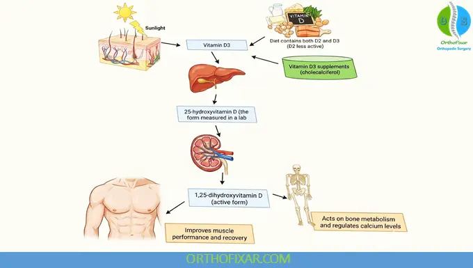
The kidneys convert 25-hydroxyvitamin D into active 1,25-dihydroxyvitamin D (calcitriol). Impaired renal function reduces active vitamin D production.
Chronic Liver Disease
The liver is responsible for vitamin D hydroxylation. Liver dysfunction can impair vitamin D metabolism.
Hypophosphatemic Disorders
Low serum phosphate levels may cause defective bone mineralization.
Examples include:
- X-linked hypophosphatemia
- Tumor-induced osteomalacia
- Fanconi syndrome
Medications
Several drugs may contribute to osteomalacia:
- Phenytoin
- Phenobarbital
- Carbamazepine
- Rifampin
- Long-term aluminum-containing antacids
Risk Factors
Common risk factors include:
- Advanced age
- Institutionalization
- Limited outdoor activity
- Veiled clothing practices
- Obesity
- Malnutrition
- Chronic kidney disease
- Gastrointestinal disorders
- Long-term anticonvulsant therapy
Symptoms of Osteomalacia
Symptoms often develop gradually and may be overlooked.
Common clinical manifestations include:
Bone Pain
Diffuse skeletal pain is one of the hallmark symptoms.
Frequently affected areas:
- Lower back
- Hips
- Pelvis
- Ribs
- Lower extremities
Muscle Weakness
Proximal muscle weakness commonly affects:
- Thigh muscles
- Shoulder girdle muscles
Patients may report:
- Difficulty climbing stairs
- Difficulty standing from a seated position
- Difficulty walking
Fatigue
Generalized weakness and chronic fatigue are common complaints.
Fragility Fractures
Softened bones are more susceptible to:
- Stress fractures
- Insufficiency fractures
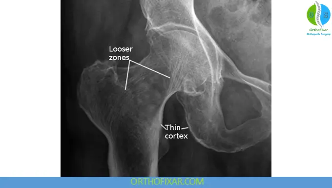
- Pseudofractures (Looser zones)
Physical Examination Findings
Healthcare providers may observe:
- Antalgic gait
- Waddling gait
- Proximal muscle weakness
- Bone tenderness
- Skeletal deformities in advanced disease
Diagnosis of Osteomalacia
Diagnosis requires a combination of clinical assessment, laboratory testing and imaging studies.
Laboratory Findings
Typical biochemical abnormalities include:
| Laboratory Test | Typical Finding |
|---|---|
| 25-Hydroxyvitamin D | Low |
| Calcium | Low or normal |
| Phosphate | Low |
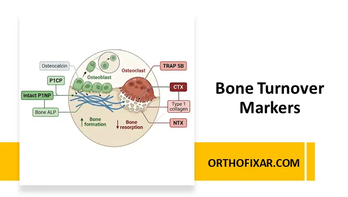
| Alkaline Phosphatase | Elevated |
| Parathyroid Hormone (PTH) | Elevated |
| 1,25-Dihydroxyvitamin D | Variable |
Elevated alkaline phosphatase is one of the most important biochemical markers.
Imaging Studies
Plain Radiographs
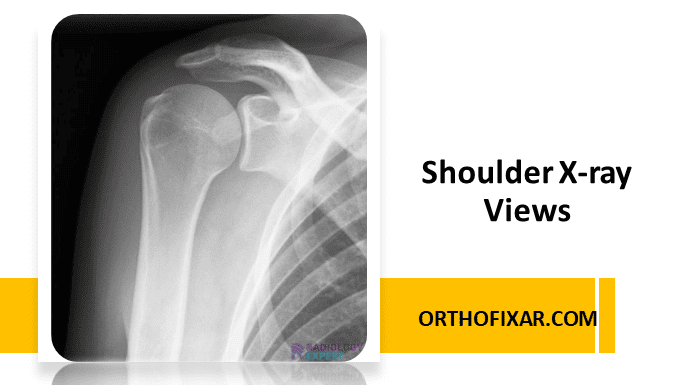
X-rays may demonstrate:
- Generalized osteopenia
- Cortical thinning
- Looser zones (pseudofractures)
Common sites include:
- Femoral neck
- Pubic rami
- Scapula
- Ribs
Bone Mineral Density (DEXA)
DEXA scanning may reveal reduced bone density but cannot distinguish osteomalacia from osteoporosis.
Bone Biopsy
Bone histomorphometry remains the definitive diagnostic test but is rarely required in routine clinical practice.
Differential Diagnosis
Conditions that may mimic osteomalacia include:
- Osteoporosis
- Multiple myeloma
- Metastatic bone disease
- Hyperparathyroidism
- Renal osteodystrophy
- Fibromyalgia
Accurate laboratory assessment is critical for differentiation.
Treatment of Osteomalacia
Treatment focuses on correcting the underlying cause and restoring normal mineralization.
Vitamin D Replacement
For vitamin D deficiency-related osteomalacia, supplementation is the cornerstone of therapy.
Common regimens include:
- Cholecalciferol (Vitamin D3)
- Ergocalciferol (Vitamin D2)
Severe deficiency may require:
- 50,000 IU weekly for 8–12 weeks
- Maintenance therapy of 800–2000 IU daily
Treatment should be individualized according to laboratory findings and patient characteristics.
Calcium Supplementation
Adequate calcium intake is necessary for effective treatment.
Recommended intake generally ranges around:
- 1000–1200 mg daily
Dietary sources include:
- Dairy products
- Fortified foods
- Leafy green vegetables
Phosphate Replacement
Patients with hypophosphatemic osteomalacia may require:
- Oral phosphate supplementation
- Active vitamin D analogs
Management of Underlying Disorders
Successful treatment often requires addressing contributing conditions such as:
- Malabsorption syndromes
- Chronic kidney disease
- Chronic liver disease
- Tumor-induced osteomalacia
Monitoring Response to Therapy
Monitoring typically includes:
- Serum calcium
- Serum phosphate
- Alkaline phosphatase
- Parathyroid hormone
- 25-hydroxyvitamin D levels
Clinical improvement often occurs within weeks, while complete skeletal recovery may take several months.
Complications of Untreated Osteomalacia
Failure to treat osteomalacia may result in:
- Chronic pain
- Progressive muscle weakness
- Fragility fractures
- Skeletal deformities
- Reduced mobility
- Increased fall risk
Prevention of Osteomalacia
Preventive measures include:
Adequate Vitamin D Intake
Adults should maintain sufficient vitamin D intake through:
- Diet
- Supplements when indicated
Safe Sun Exposure
Regular sunlight exposure supports endogenous vitamin D production.
Adequate Calcium Consumption
Balanced dietary calcium intake is essential for bone health.
Screening High-Risk Populations
Early detection is recommended for:
- Older adults
- Patients with malabsorption
- Chronic kidney disease patients
- Long-term anticonvulsant users
Osteomalacia vs Osteoporosis
| Feature | Osteomalacia | Osteoporosis |
|---|---|---|
| Primary Defect | Poor mineralization | Reduced bone mass |
| Bone Quality | Soft bones | Fragile bones |
| Vitamin D Deficiency | Common | May coexist |
| Alkaline Phosphatase | Elevated | Usually normal |
| Treatment | Correct mineral deficiency | Antiresorptive/anabolic therapy |
Prognosis
The prognosis of osteomalacia is generally excellent when diagnosed early and treated appropriately.
Most patients experience:
- Reduced bone pain
- Improved muscle strength
- Enhanced mobility
- Decreased fracture risk
Delayed diagnosis may result in irreversible skeletal deformities and prolonged disability.
Key Takeaways
- Osteomalacia is a disorder of defective bone mineralization in adults.
- Vitamin D deficiency is the most common cause worldwide.
- Symptoms include bone pain, muscle weakness, fatigue, and fractures.
- Laboratory findings typically show low vitamin D, elevated alkaline phosphatase, and secondary hyperparathyroidism.
- Treatment focuses on vitamin D, calcium, phosphate replacement, and management of underlying causes.
- Early diagnosis significantly improves outcomes and prevents complications.
References & More
- Zimmerman L, Anastasopoulou C, McKeon B. Osteomalacia. [Updated 2024 Sep 2]. In: StatPearls [Internet]. Treasure Island (FL): StatPearls Publishing; 2026 Jan-. Available from: https://www.ncbi.nlm.nih.gov/books/NBK551616/
- Zimmerman L, Anastasopoulou C, McKeon B. Osteomalacia. 2024 Sep 2. In: StatPearls [Internet]. Treasure Island (FL): StatPearls Publishing; 2026 Jan–. PMID: 31869080. Pubmed