What is Hypocalcaemia?
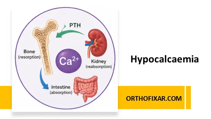
Hypocalcaemia is defined as a decreased concentration of serum calcium, usually a corrected total serum calcium below 2.1 mmol/L (8.5 mg/dL) or ionized calcium below 1.1 mmol/L, depending on laboratory reference ranges. Calcium is essential for neuromuscular transmission, muscle contraction, blood coagulation, hormone secretion, and cardiac electrophysiology.
The body maintains serum calcium within a narrow range through the coordinated actions of:
- Parathyroid hormone (PTH)
- Vitamin D (calcitriol)
- Kidneys
- Bones
- Gastrointestinal tract
Symptomatic hypocalcaemia is relatively uncommon because these homeostatic mechanisms are highly efficient. However, when hypocalcaemia develops rapidly or becomes severe, it represents a medical emergency requiring immediate treatment.
Calcium Homeostasis
Role of Parathyroid Hormone (PTH)
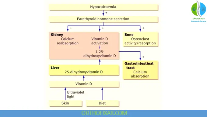
PTH increases serum calcium by:
- Stimulating bone resorption
- Increasing renal calcium reabsorption
- Promoting renal activation of vitamin D
- Enhancing intestinal calcium absorption indirectly via calcitriol
Role of Vitamin D
Active vitamin D (1,25-dihydroxyvitamin D):
- Increases intestinal calcium absorption
- Enhances phosphate absorption
- Supports bone mineralization
Role of the Kidneys
The kidneys:
- Activate vitamin D
- Reabsorb calcium
- Excrete phosphate
- Respond to PTH stimulation
Failure of any of these mechanisms may lead to hypocalcaemia.
See Also: Bone Formation & Development
Causes of Hypocalcaemia
Hypoparathyroidism
The most important cause of persistent hypocalcaemia is hypoparathyroidism.
Common causes include:
- Post-thyroidectomy or parathyroidectomy
- Autoimmune hypoparathyroidism
- Congenital absence of parathyroid glands (DiGeorge syndrome)
- Iron overload (haemochromatosis)
- Magnesium deficiency
- Activating mutations of the calcium-sensing receptor (CaSR)
Laboratory findings include:
- Low calcium
- High phosphate
- Low or absent PTH
Vitamin D Deficiency
Vitamin D deficiency reduces intestinal calcium absorption.
Causes include:
- Poor dietary intake
- Limited sunlight exposure
- Malabsorption
- Chronic liver disease
- Chronic kidney disease
Chronic Kidney Disease (CKD)
CKD causes hypocalcaemia through:
- Reduced calcitriol production
- Phosphate retention
- Secondary hyperparathyroidism
- Impaired calcium absorption
Magnesium Deficiency
Hypomagnesaemia suppresses:
- PTH secretion
- Peripheral response to PTH
Hypocalcaemia may not correct until magnesium deficiency is treated.
Pseudohypoparathyroidism
A rare inherited disorder characterized by resistance to PTH.
Features include:
- Elevated PTH
- Low calcium
- High phosphate
- End-organ resistance to PTH
Type Ia
Associated with Albright hereditary osteodystrophy, characterized by:
- Short stature
- Round face
- Obesity
- Short fourth and fifth metacarpals
- Developmental delay in some patients
Type Ib
Primarily presents with biochemical abnormalities without obvious skeletal features.
Other Causes
- Acute pancreatitis
- Massive blood transfusion (citrate toxicity)
- Tumor lysis syndrome
- Rhabdomyolysis
- Sepsis
- Certain medications:
- Bisphosphonates
- Denosumab
- Cinacalcet
- Loop diuretics
- Phenytoin
Risk Factors
Patients at increased risk include:
- Recent thyroid or parathyroid surgery
- Chronic kidney disease
- Malnutrition
- Elderly individuals
- Malabsorption syndromes
- Vitamin D deficiency
- Alcohol dependence
- Critically ill patients
Pathophysiology
Reduced extracellular calcium lowers the threshold for neuronal depolarization.
This causes:
- Increased neuromuscular excitability
- Tetany
- Muscle spasms
- Seizures
- Cardiac electrical instability
Chronic hypocalcaemia may also affect:
- Bone metabolism
- Tooth development
- Lens transparency
- Basal ganglia
Clinical Features
Clinical manifestations depend on:
- Severity
- Rate of onset
- Duration
Acute hypocalcaemia generally causes more severe symptoms than chronic disease.
Neuromuscular Symptoms
- Perioral numbness
- Tingling of fingers and toes
- Muscle cramps
- Tetany
- Carpopedal spasm
- Generalized muscle stiffness
- Hyperreflexia
Neurological Symptoms
- Anxiety
- Irritability
- Fatigue
- Confusion
- Seizures
- Cognitive impairment
Cardiovascular Manifestations
- Hypotension
- Heart failure (rare)
- Ventricular arrhythmias
- Prolonged QT interval
- Torsades de pointes (rare)
Respiratory Manifestations
- Laryngospasm
- Bronchospasm
- Respiratory distress
Classical Clinical Signs
Chvostek Sign
Chvostek sign is elicited by tapping over the facial nerve anterior to the ear.
A positive sign produces:
- Facial muscle twitching
- Ipsilateral facial contraction
It indicates increased neuromuscular excitability but lacks specificity.
Trousseau Sign
Trousseau sign is more sensitive than Chvostek sign.
It is produced by inflating a blood pressure cuff above systolic pressure for approximately 3 minutes.
A positive response causes:
- Carpal spasm
- Wrist flexion
- MCP flexion
- Finger extension
- Thumb adduction
ECG Changes in Hypocalcaemia
The classic ECG finding is:
- Prolonged QT interval
Other possible findings include:
- ST-segment prolongation
- Ventricular arrhythmias (rare)
Investigations
Initial Laboratory Tests
Essential investigations include:
- Corrected serum calcium
- Ionized calcium
- Serum albumin
- Serum phosphate
- Serum magnesium
- Intact PTH
- 25-hydroxyvitamin D
- Renal function tests
- Liver function tests
Additional Tests
Depending on clinical suspicion:
- Calcium-sensing receptor mutation analysis
- Genetic testing
- Urinary cyclic AMP response to PTH
- Autoimmune screening
- Iron studies
- Urinary calcium excretion
Imaging
Imaging may reveal:
- Basal ganglia calcification
- Soft tissue calcification
- Nephrocalcinosis
- Nephrolithiasis
Radiographs may also identify skeletal abnormalities associated with congenital disorders.
Differential Diagnosis
Conditions that may mimic hypocalcaemia include:
- Hyperventilation syndrome
- Hypomagnesaemia
- Epilepsy
- Tetanus
- Dystonia
- Conversion disorder
- Hypoglycaemia
Treatment of Hypocalcaemia
Treatment depends on symptom severity and the underlying cause.
Acute Symptomatic Hypocalcaemia
Acute symptomatic hypocalcaemia is a medical emergency.
Management includes:
- Continuous cardiac monitoring
- Intravenous calcium gluconate (preferred)
- Slow IV administration with ECG monitoring
- Correction of hypomagnesaemia
- Identification and treatment of the underlying cause
Calcium chloride contains more elemental calcium but is generally reserved for central venous administration because of its risk of tissue necrosis if extravasation occurs.
Chronic Hypocalcaemia
Long-term treatment usually includes:
- Oral calcium supplements
- Calcitriol
- Vitamin D replacement (when deficient)
- Magnesium replacement (if deficient)
Patients require regular monitoring to avoid:
- Hypercalcaemia
- Hypercalciuria
- Nephrolithiasis
- Nephrocalcinosis
Management of Chronic Hypoparathyroidism
Patients with chronic hypoparathyroidism may benefit from:
- Oral calcium
- Active vitamin D analogues
- Recombinant PTH therapy (selected patients where available)
The treatment goal is maintaining serum calcium in the low-normal range while minimizing urinary calcium excretion.
Complications
Untreated hypocalcaemia may lead to:
- Seizures
- Cardiac arrhythmias
- Laryngospasm
- Heart failure
- Cataracts
- Basal ganglia calcification
- Neurocognitive dysfunction
- Chronic renal complications from overtreatment
Prognosis
The prognosis depends on the underlying cause.
Excellent outcomes are expected when:
- Vitamin D deficiency is corrected
- Magnesium deficiency is treated
- Acute postoperative hypocalcaemia is recognized early
Patients with permanent hypoparathyroidism require lifelong follow-up and biochemical monitoring.
Prevention
Preventive measures include:
- Adequate dietary calcium intake
- Sufficient vitamin D levels
- Monitoring after thyroid and parathyroid surgery
- Early treatment of magnesium deficiency
- Appropriate management of chronic kidney disease
Key Points
- Hypocalcaemia is defined by low serum or ionized calcium.
- The most common major causes are hypoparathyroidism, vitamin D deficiency, and chronic kidney disease.
- Acute symptomatic hypocalcaemia is a medical emergency requiring intravenous calcium gluconate.
- Chvostek sign and Trousseau sign are classic clinical findings of neuromuscular excitability.
- ECG typically demonstrates QT interval prolongation.
- Always measure PTH, magnesium, phosphate, vitamin D, and renal function when evaluating hypocalcaemia.
- Chronic treatment focuses on oral calcium, vitamin D metabolites, and correction of the underlying disorder while avoiding hypercalciuria.
Frequently Asked Questions (FAQ)
What is the most common cause of hypocalcaemia?
The most common causes include vitamin D deficiency, hypoparathyroidism, and chronic kidney disease, with prevalence varying by clinical setting.
Why does hypocalcaemia prolong the QT interval?
Low extracellular calcium prolongs phase 2 (plateau phase) of the cardiac action potential, resulting in QT interval prolongation.
Which clinical sign is more sensitive for hypocalcaemia?
Trousseau sign is generally considered more sensitive and specific than Chvostek sign.
Why should magnesium be checked in hypocalcaemia?
Magnesium deficiency impairs both PTH secretion and its peripheral action. Hypocalcaemia often cannot be corrected until magnesium levels are restored.
What is the emergency treatment for severe hypocalcaemia?
Symptomatic severe hypocalcaemia requires prompt intravenous calcium (typically calcium gluconate), ECG monitoring, correction of hypomagnesaemia if present, and treatment of the underlying cause.
References & More
- Goyal A, Anastasopoulou C, Ngu M, et al. Hypocalcemia. [Updated 2023 Oct 15]. In: StatPearls [Internet]. Treasure Island (FL): StatPearls Publishing; 2026 Jan-. Available from: https://www.ncbi.nlm.nih.gov/books/NBK430912/
- Cooper MS, Gittoes NJ. Diagnosis and management of hypocalcaemia. BMJ. 2008 Jun 7;336(7656):1298-302. doi: 10.1136/bmj.39582.589433.BE. Erratum in: BMJ. 2008 Jun 28;336(7659): doi: 10.1136/bmj.a334. PMID: 18535072; PMCID: PMC2413335. Link
- Schafer AL, Shoback DM. Hypocalcemia: Diagnosis and Treatment. [Updated 2016 Jan 3]. In: Feingold KR, Adler RA, Ahmed SF, et al., editors. Endotext [Internet]. South Dartmouth (MA): MDText.com, Inc.; 2000-. Available from: https://www.ncbi.nlm.nih.gov/books/NBK279022/