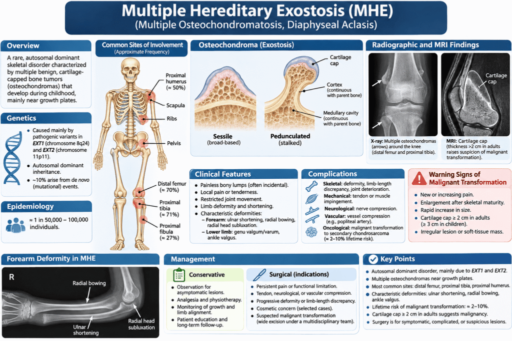
Multiple hereditary exostosis (MHE), also known as hereditary multiple exostoses, hereditary multiple osteochondromas, multiple osteochondromatosis, or diaphyseal aclasis, is a rare inherited skeletal disorder characterized by the development of multiple benign, cartilage-capped bone tumors called osteochondromas.
These lesions typically develop during childhood near active growth plates and may result in progressive skeletal deformities, limb shortening, restricted joint movement, neurovascular compression, and, occasionally, malignant transformation into secondary chondrosarcoma.
What Is Multiple Hereditary Exostosis?
Multiple hereditary exostosis is a genetic musculoskeletal disorder in which numerous osteochondromas develop on the external surfaces of bones formed through endochondral ossification.
An osteochondroma consists of three principal components:
- An outer cortical bone layer.
- An internal medullary cavity continuous with that of the parent bone.
- An overlying hyaline cartilage cap.
Unlike solitary osteochondroma, multiple hereditary exostosis represents a generalized skeletal disorder with an inherited genetic predisposition.
Epidemiology
Hereditary multiple exostoses is an uncommon skeletal condition with an estimated frequency of approximately 1 in 50,000 to 100,000 individuals.
The main epidemiological characteristics include:
- Onset during childhood.
- Median age at diagnosis of approximately three years.
- Most affected individuals are diagnosed before 12 years of age.
- Progressive development of osteochondromas during skeletal growth.
- Cessation of lesion growth in most cases following skeletal maturity.
The clinical severity varies considerably among affected individuals, even within the same family. [1,2]
Etiology and Genetics of Multiple Hereditary Exostosis
Multiple hereditary exostosis is primarily caused by pathogenic variants in the EXT1 and EXT2 tumor suppressor genes.
| Gene | Chromosomal location | Function |
|---|---|---|
| EXT1 | Chromosome 8q24 | Heparan sulfate synthesis |
| EXT2 | Chromosome 11p11 | Heparan sulfate synthesis |
Both genes encode glycosyltransferases involved in the biosynthesis of heparan sulfate, which plays an essential role in regulating cartilage development and skeletal growth.
Although an EXT3 locus on chromosome 19 was historically proposed, EXT1 and EXT2 are the established causative genes.
Inheritance Pattern
Multiple hereditary exostosis follows an autosomal dominant pattern of inheritance.
- Approximately 90% of patients have an affected parent.
- Approximately 10% develop the condition through de novo pathogenic variants.
- Each child of an affected parent has a 50% probability of inheriting the disease-causing variant.
Genotype–Phenotype Correlation
Patients with EXT1 pathogenic variants tend to have more extensive skeletal involvement and more severe deformities than patients with EXT2 variants. However, considerable clinical variability exists, and genotype alone cannot reliably predict individual outcomes. [1,3]
See Also: Osteochondroma: Comprehensive Medical Overview
Pathophysiology
The underlying mechanism of multiple hereditary exostosis involves defective heparan sulfate production and disruption of the signaling pathways responsible for normal skeletal development.
Heparan sulfate regulates several molecular pathways, including those involving bone morphogenetic proteins (BMPs), fibroblast growth factors (FGFs), and Hedgehog signaling.
A deficiency of heparan sulfate interferes with normal chondrocyte differentiation and growth plate organization.
The development of osteochondromas can be summarized as follows:
- Pathogenic variants in EXT1 or EXT2 impair heparan sulfate synthesis.
- Abnormal signaling occurs within the growth plate and adjacent perichondrium.
- Ectopic cartilaginous tissue develops near the peripheral growth plate.
- The cartilaginous tissue undergoes endochondral ossification.
- A cartilage-capped osseous projection develops.
- Continued longitudinal bone growth causes the lesion to become relatively displaced from the physis.
Additional somatic genetic alterations, consistent with the two-hit hypothesis, may contribute to osteochondroma formation.
Morphological Types of Osteochondromas
Osteochondromas are classified according to their morphological appearance into two main types:
Sessile Osteochondroma
A broad-based osseous projection directly attached to the underlying bone.
Pedunculated Osteochondroma
An osseous projection connected to the parent bone by a relatively narrow stalk, typically directed away from the adjacent joint. [3,4,5]
Common Anatomical Sites
Multiple hereditary exostosis predominantly affects the metaphyseal regions of long bones, particularly around the knee joint.
Frequently affected anatomical sites include:
| Anatomical site | Approximate frequency |
|---|---|
| Distal femur | 70% |
| Proximal tibia | 71% |
| Humerus | 50% |
| Radius and ulna | 30% |
| Proximal fibula | 27% |
Other sites of involvement include the pelvis, scapula, ribs, ankle, vertebral column, hands, and feet.
The anatomical distribution and number of osteochondromas vary between patients and published studies. [3]
Clinical Presentation of Multiple Hereditary Exostosis
Multiple hereditary exostosis usually presents during childhood with one or more palpable, firm bony masses.
Although many osteochondromas remain asymptomatic, clinical manifestations depend on their location, size, and effects on surrounding anatomical structures.
Symptoms
Common presenting symptoms include:
- Painless palpable bony masses.
- Localized pain and tenderness.
- Restricted joint range of motion.
- Progressive limb deformity.
- Limb-length discrepancy.
- Short stature.
- Mechanical irritation of surrounding muscles and tendons.
- Neurological symptoms secondary to nerve compression.
- Vascular symptoms caused by compression of adjacent blood vessels.
Skeletal Deformities
Disturbance of physeal growth and differential growth between paired bones can result in significant skeletal abnormalities.
Forearm Deformities
Forearm involvement is a characteristic manifestation of multiple hereditary exostosis.
Progressive shortening of the ulna relative to the radius may result in:
- Ulnar shortening.
- Radial bowing.
- Increased inclination of the distal radial articular surface.
- Ulnar deviation of the wrist.
- Carpal translation.
- Radial head subluxation or dislocation.
- Restricted forearm pronation and supination.
Severe deformities can significantly impair upper-limb function.
Lower-Limb Deformities
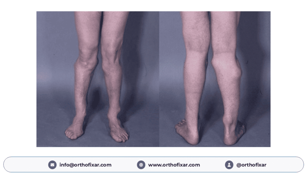
Osteochondromas affecting the lower extremities may cause:
- Genu valgum.
- Genu varum.
- Coxa valga.
- Ankle valgus.
- Limb-length discrepancy.
- Restricted joint movement.
Relative fibular shortening may contribute to progressive ankle valgus, altered joint mechanics, and premature degenerative changes. [6,7]
Complications of Multiple Hereditary Exostosis
The complications of multiple hereditary exostosis may be classified into mechanical, skeletal, neurological, vascular, and oncological complications.
Mechanical Complications
- Tendon impingement.
- Muscle irritation.
- Reactive bursitis.
- Restricted joint mobility.
- Fracture through the stalk of a pedunculated osteochondroma.
- Chronic musculoskeletal pain.
Neurological Complications
Compression of adjacent neural structures may result in peripheral neuropathy, sensory disturbances, muscle weakness, or motor dysfunction.
Spinal osteochondromas can occasionally produce nerve root or spinal cord compression, leading to radiculopathy or myelopathy.
Vascular Complications
Vascular complications are uncommon but may include:
- Arterial compression.
- Pseudoaneurysm formation.
- Arterial thrombosis.
- Venous obstruction.
- Limb ischemia.
The popliteal vessels are particularly vulnerable because of their proximity to osteochondromas around the knee.
New neurological deficits or signs of acute vascular compromise require urgent medical assessment.
Malignant Transformation in Multiple Hereditary Exostosis
The most serious long-term complication of multiple hereditary exostosis is malignant transformation of an osteochondroma into secondary peripheral chondrosarcoma.
The estimated lifetime risk of malignant transformation is approximately 2–10%. A systematic review published in 2018 reported an average transformation rate of approximately 4%.
Malignant transformation occurs predominantly during adulthood, particularly between 20 and 40 years of age.
Common Sites of Malignant Transformation
- Pelvis.
- Proximal femur.
- Scapula.
- Proximal humerus.
- Ribs.
Warning Signs of Malignant Transformation
The following findings should raise suspicion of secondary chondrosarcoma:
- New or progressively increasing pain.
- Enlargement of a previously stable osteochondroma after skeletal maturity.
- Rapid increase in lesion size.
- Irregularity of the lesion surface.
- Destruction of adjacent bone.
- Development of an associated soft-tissue mass.
- Abnormal thickening of the cartilage cap.
A cartilage cap measuring approximately 2 cm or more in a skeletally mature patient is particularly concerning and requires further investigation. However, cartilage cap thickness alone does not establish a diagnosis of malignancy. [1,8,9]
Diagnosis of Multiple Hereditary Exostosis
The diagnosis is primarily established through clinical examination, characteristic radiographic findings, and, when appropriate, molecular genetic testing.
Clinical Examination
Clinical assessment should include:
- Family history of osteochondromas or skeletal deformities.
- Examination of palpable bony masses.
- Assessment of limb alignment.
- Measurement of limb lengths.
- Evaluation of joint range of motion.
- Neurological and vascular examination.
- Assessment of pain and functional limitations.
Radiographic Investigations
Plain Radiographs
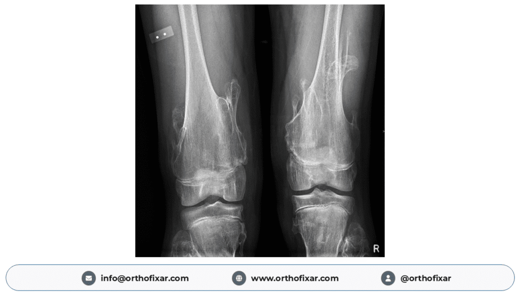
Plain radiography is the initial imaging modality for diagnosing multiple hereditary exostosis.
Characteristic radiographic findings include:
- Multiple osseous projections arising near the metaphysis.
- Continuity of the lesion cortex with the parent bone cortex.
- Continuity of the medullary cavity with that of the underlying bone.
- Sessile or pedunculated morphology.
- Pedunculated lesions typically projecting away from the adjacent joint.
- Variable calcification of the cartilage cap.
- Associated skeletal deformities.
Cortical and medullary continuity with the underlying bone is the hallmark radiographic feature of an osteochondroma.
Standing long-leg alignment radiographs may be required to assess lower-limb deformities and mechanical axis deviation.
Forearm and elbow radiographs are useful when radial bowing, ulnar shortening, or restriction of forearm rotation is present.
Computed Tomography (CT)
CT is particularly useful for evaluating osteochondromas in anatomically complex areas, including:
- Pelvis.
- Scapula.
- Vertebral column.
- Ribs.
CT provides detailed information regarding osseous anatomy, cortical continuity, and relationships with adjacent structures.
Magnetic Resonance Imaging (MRI)
MRI is the preferred imaging modality for evaluating the cartilage cap and suspected malignant transformation.
MRI is also useful for identifying:
- Soft-tissue involvement.
- Neurovascular compression.
- Reactive bursitis.
- Spinal cord compression.
- Associated soft-tissue masses.
Cartilage Cap Thickness
The cartilage cap characteristically demonstrates low to intermediate signal intensity on T1-weighted images and high signal intensity on T2-weighted images.
A cartilage cap thickness greater than 2 cm in adults or greater than 3 cm in children may indicate malignant transformation and warrants specialist evaluation in the appropriate clinical context.
Children may normally have thicker cartilage caps than adults because of ongoing skeletal growth.
Ultrasonography
Ultrasound may assist in assessing the cartilage cap of superficial osteochondromas, identifying reactive bursae, and investigating vascular complications. [2,4,9,10]
Genetic Testing
Molecular genetic testing can identify pathogenic variants in EXT1 and EXT2.
Genetic testing is particularly useful in patients with atypical findings, uncertain diagnoses, or a need for genetic counseling and family assessment.
Differential Diagnosis
The differential diagnosis of multiple hereditary exostosis includes several disorders associated with osseous or cartilaginous lesions.
| Condition | Distinguishing characteristics |
|---|---|
| Solitary osteochondroma | Single osteochondral lesion |
| Ollier disease | Multiple intramedullary enchondromas |
| Maffucci syndrome | Enchondromas associated with vascular anomalies |
| Dysplasia epiphysealis hemimelica (Trevor disease) | Predominantly asymmetric epiphyseal osteochondral overgrowth |
| Metachondromatosis | Multiple exostosis-like lesions and enchondromas |
| Bizarre parosteal osteochondromatous proliferation (Nora lesion) | Surface lesion usually lacking typical medullary continuity |
| Secondary chondrosarcoma | Suspicious post-maturity growth, pain, or cartilage cap thickening |
Treatment of Multiple Hereditary Exostosis
The treatment of multiple hereditary exostosis is individualized according to the patient’s symptoms, age, skeletal maturity, deformity severity, and risk of complications.
Routine removal of every osteochondroma is not recommended.
Conservative Treatment
Asymptomatic osteochondromas generally do not require surgical intervention.
Conservative management may include:
- Regular clinical assessment.
- Observation of asymptomatic lesions.
- Appropriate analgesia.
- Physiotherapy when indicated.
- Monitoring skeletal growth and limb alignment.
- Patient education regarding malignant transformation.
Surgical Treatment
Surgical intervention is indicated when osteochondromas cause significant symptoms, functional impairment, progressive deformity, or other complications.
Indications for Surgery
- Persistent pain.
- Restricted joint movement.
- Symptomatic tendon or muscle impingement.
- Neurological compression.
- Vascular compression.
- Progressive skeletal deformity.
- Significant cosmetic concerns after individualized assessment.
- Suspected malignant transformation.
Osteochondroma Excision
Symptomatic osteochondromas may be treated with complete surgical excision at their base.
The entire cartilage cap and its perichondrial covering should be removed to minimize recurrence. Care is required to preserve adjacent growth plates, nerves, blood vessels, and other important structures.
Correction of Forearm Deformity
Surgical options for significant forearm deformities include:
- Excision of symptomatic osteochondromas.
- Ulnar lengthening using distraction osteogenesis.
- Corrective radial osteotomy.
- Selected procedures addressing radial head instability.
Correction of Lower-Limb Deformity
Treatment options include:
- Guided growth using temporary hemiepiphysiodesis.
- Corrective osteotomy.
- Epiphysiodesis for selected limb-length discrepancies.
- Limb-lengthening procedures in selected cases.
Early correction of progressive ankle valgus during childhood may help preserve long-term joint function.
Management of Malignant Transformation
Patients with suspected secondary chondrosarcoma should be referred to a specialist orthopedic oncology center.
Management may involve:
- MRI for local tumor assessment.
- Appropriate staging investigations.
- Biopsy when indicated and planned by the treating sarcoma team.
- Multidisciplinary assessment.
- Oncologically appropriate surgical resection, often with wide margins.
Unplanned excision of a lesion suspected of malignant transformation should be avoided. [1,6,7,11]
Follow-Up and Prognosis
The prognosis of multiple hereditary exostosis is generally favorable because most osteochondromas are benign.
Nevertheless, long-term orthopedic follow-up is important to identify progressive deformities, neurological or vascular complications, and potential malignant transformation.
During childhood, follow-up primarily focuses on skeletal growth, limb alignment, and preservation of joint function.
During adulthood, particular attention should be given to newly painful or enlarging lesions, especially those involving the pelvis and scapula.
There is currently no universally accepted imaging surveillance schedule. Imaging should be individualized according to symptoms, lesion location, clinical findings, and specialist recommendations.
Genetic counseling should be offered to affected individuals and their families.
Multiple Hereditary Exostosis: Key Orthopedic Points
- Multiple hereditary exostosis is an autosomal dominant skeletal disorder.
- EXT1 and EXT2 are the principal causative genes.
- Osteochondromas usually arise near the metaphyses of growing bones.
- Cortical and medullary continuity is the characteristic radiographic finding.
- Ulnar shortening and ankle valgus are important orthopedic manifestations.
- Malignant transformation occurs in approximately 2–10% of affected individuals over their lifetime.
- New pain or lesion growth after skeletal maturity requires investigation.
- A cartilage cap measuring 2 cm or more in an adult raises suspicion of secondary chondrosarcoma.
- Surgery is reserved for appropriately selected symptomatic or complicated lesions.
Frequently Asked Questions
Is multiple hereditary exostosis cancerous?
No. Most osteochondromas are benign. However, affected individuals have an increased lifetime risk of developing secondary chondrosarcoma.
What genes cause multiple hereditary exostosis?
The condition is primarily caused by pathogenic variants in the EXT1 gene on chromosome 8 and the EXT2 gene on chromosome 11.
Do osteochondromas stop growing after puberty?
Osteochondromas generally stop enlarging after skeletal maturity. Continued or renewed growth in adulthood requires further evaluation.
What is the most common site of multiple hereditary exostosis?
The knee region is among the most commonly affected areas, especially the distal femur and proximal tibia.
What cartilage cap thickness suggests malignancy?
A cartilage cap measuring approximately 2 cm or more in a skeletally mature patient raises suspicion of malignant transformation.
Does multiple hereditary exostosis require surgery?
Not all patients require surgery. Surgical intervention is considered for symptomatic lesions, progressive deformities, neurovascular compression, or suspected malignancy.
Can multiple hereditary exostosis be cured?
There is currently no established treatment that corrects the underlying genetic disorder. Treatment focuses on managing symptoms, correcting deformities, and addressing complications.
References
- Hereditary Multiple Osteochondromas. GeneReviews. NCBI Bookshelf. Updated January 2026. https://www.ncbi.nlm.nih.gov/books/NBK1235/
- Hereditary multiple exostoses: an educational review. 2025. PMID: 39982564. https://pubmed.ncbi.nlm.nih.gov/39982564/
- Hereditary Multiple Exostoses—A Review of the Molecular Background, Diagnostics, and Potential Therapeutic Strategies. 2021. https://pmc.ncbi.nlm.nih.gov/articles/PMC8704583/
- Osteochondromas: An Updated Review of Epidemiology, Pathogenesis, Clinical Presentation, Radiological Features and Treatment Options. 2021. PMID: 33622860. https://pubmed.ncbi.nlm.nih.gov/33622860/
- D’Arienzo A, et al. Hereditary Multiple Exostoses: Current Insights. Orthop Res Rev. 2019;11:199–211. https://pubmed.ncbi.nlm.nih.gov/31853203/
- Forearm deformity in patients with hereditary multiple exostoses: factors associated with range of motion and radial head dislocation. 2013. https://pubmed.ncbi.nlm.nih.gov/24005199/
- Whalen MJ, van Bosse HJP. Management of Lower Extremity Deformity in Children with Hereditary Multiple Exostoses. 2025. https://pubmed.ncbi.nlm.nih.gov/41158744/
- Fei L, Ngoh C, Porter DE. Chondrosarcoma transformation in hereditary multiple exostoses: A systematic review. J Bone Oncol. 2018;13:114–122. https://pubmed.ncbi.nlm.nih.gov/30591865/
- Bernard SA, et al. Improved differentiation of benign osteochondromas from secondary chondrosarcomas with standardized measurement of cartilage cap at CT and MR imaging. Radiology. 2010;255(3):857–865. https://pubmed.ncbi.nlm.nih.gov/20392983/
- Multimodality imaging features of hereditary multiple exostoses. 2013. https://pubmed.ncbi.nlm.nih.gov/24004486/
- Strauss SJ, et al. Bone sarcomas: ESMO-EURACAN-GENTURIS-ERN PaedCan Clinical Practice Guideline for diagnosis, treatment and follow-up. Ann Oncol. 2021;32(12):1520–1536. https://pubmed.ncbi.nlm.nih.gov/34500044/