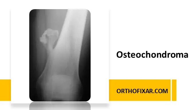
Osteochondroma is the most common benign bone tumor, accounting for approximately 35–50% of all benign bone tumors and 8–15% of all bone tumors overall (Schmale et al., 2001). It arises from the growth plate (physis) and typically presents as an exophytic lesion covered by cartilage.
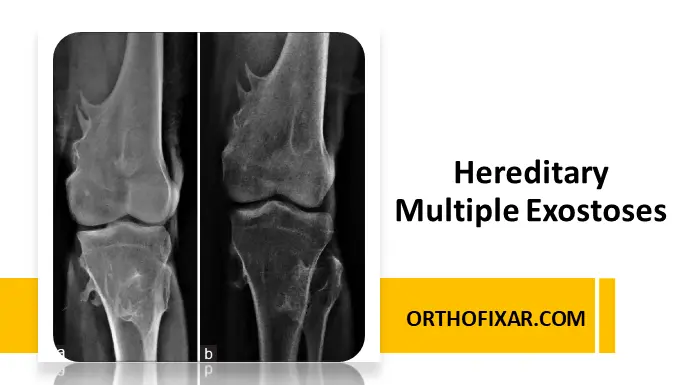
Osteochondromas can occur as a solitary lesion or as multiple lesions in the context of hereditary multiple exostoses (HME), an autosomal dominant disorder.
Epidemiology
- Age: Most commonly diagnosed in children and adolescents, often between ages 10–20.
- Gender: Slight male predominance (male-to-female ratio 1.5:1).
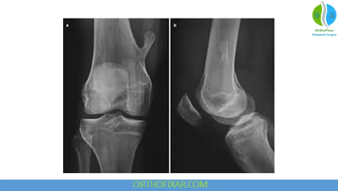
- Sites: Frequently occurs in the metaphyseal region of long bones, particularly the distal femur, proximal tibia, and proximal humerus.
Etiology and Pathophysiology
Osteochondromas develop due to aberrant cartilage growth at the physis, leading to an outward bony projection covered with a cartilage cap. Mutations in EXT1 and EXT2 genes are implicated, particularly in hereditary multiple exostoses.
The cartilage cap thickness is clinically significant: lesions with cap thickness >2 cm in adults warrant suspicion for malignant transformation into chondrosarcoma.
See Also: Hereditary Multiple Exostoses (HME): Causes, Symptoms & Treatment
Clinical Presentation
Most osteochondromas are asymptomatic and discovered incidentally. Symptomatic cases may present with:
- Palpable, painless mass near a joint
- Mechanical irritation or compression of nearby muscles, tendons, or neurovascular structures
- Limb deformity, especially in multiple osteochondromas
- Rarely, pain due to fracture of the stalk or malignant transformation
Diagnostic Evaluation
Imaging Studies:
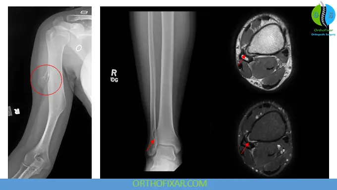
- X-ray: First-line imaging; shows sessile or pedunculated bony projection with continuous marrow and cortical continuity.
- CT Scan: Useful for complex anatomy or pre-surgical planning.
- MRI: Best for evaluating cartilage cap thickness and differentiating benign from malignant transformation.
Histopathology:
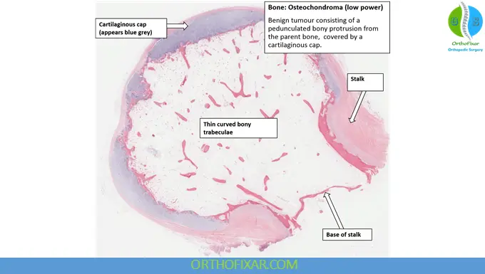
- Confirms the presence of a cartilage cap and underlying bone.
- Malignant transformation shows increased cellularity and atypia in the cartilage cap.
Osteochondroma Treatment
Conservative Management:
- Indicated for asymptomatic lesions.
- Regular follow-up to monitor growth, particularly in children.
Surgical Management:
- Osteochondroma surgery removal is recommended for symptomatic lesions causing pain, neurovascular compromise, or suspected malignancy.
- Complete excision including the cartilage cap is essential to prevent recurrence.
Follow-up:
- Long-term follow-up is advised for patients with HME due to increased risk of malignant transformation.
Complications
- Mechanical symptoms (pain, limited range of motion)
- Fracture of the osteochondroma stalk
- Neurovascular compression
- Rare malignant transformation (<1% in solitary lesions; up to 5–25% in HME)
Prognosis
Solitary osteochondromas generally have an excellent prognosis after surgical excision. Recurrence is rare if the lesion is fully excised. Hereditary multiple exostoses require closer monitoring due to a higher risk of complications and malignancy.
Key Takeaways for Medical Professionals
- Osteochondroma is a benign cartilage-capped bony projection from the metaphysis.
- Most patients are asymptomatic; symptomatic lesions require careful evaluation.
- Imaging with X-ray and MRI is essential for diagnosis and surgical planning.
- Surgical excision is curative for symptomatic lesions, while malignant transformation remains rare.
References:
- Alabdullrahman LW, Mabrouk A, Byerly DW. Osteochondroma. [Updated 2024 Feb 26]. In: StatPearls [Internet]. Treasure Island (FL): StatPearls Publishing; 2026 Jan-. Available from: https://www.ncbi.nlm.nih.gov/books/NBK544296/
- Alabdullrahman LW, Mabrouk A, Byerly DW. Osteochondroma. 2024 Feb 26. In: StatPearls [Internet]. Treasure Island (FL): StatPearls Publishing; 2026 Jan–. PMID: 31335016. Pubmed