Diastrophic dysplasia (DTD) is a rare genetic skeletal dysplasia characterized by disproportionate short stature, congenital joint contractures, distinctive hand and foot deformities, and abnormalities of cartilage and bone development. It is inherited in an autosomal recessive pattern and is caused by pathogenic variants in the SLC26A2 gene, which encodes a sulfate transporter essential for normal cartilage formation.
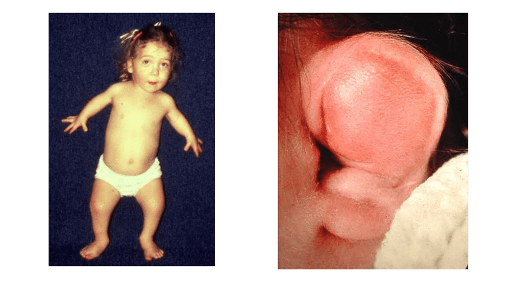
The condition is typically recognized at birth or during infancy because of characteristic physical findings, including hitchhiker thumbs, clubfoot, swelling and deformity of the external ears, and, in some patients, cleft palate. As affected individuals grow, additional musculoskeletal problems may develop, including scoliosis, hip and knee contractures, and cervical spine abnormalities.
Early diagnosis and coordinated multidisciplinary care are important to preserve joint mobility, improve walking ability, manage spinal complications, and support long-term function. Although diastrophic dysplasia is a lifelong condition, appropriate orthopedic management, rehabilitation, and surveillance can improve quality of life.
What Is Diastrophic Dysplasia?
Diastrophic dysplasia is a congenital skeletal disorder belonging to the group of autosomal recessive SLC26A2-related skeletal dysplasias. It primarily affects the development of cartilage, which is essential for normal bone growth and the formation of many joints.
The disorder is associated with short-limbed disproportionate stature, congenital deformities of the extremities, joint contractures, and variable spinal abnormalities. Its severity differs among individuals, but the musculoskeletal manifestations can be substantial and may require repeated orthopedic interventions.
Diastrophic dysplasia is also known as diastrophic dwarfism. It should be distinguished from other skeletal dysplasias, including achondroplasia and other conditions associated with pathogenic variants in SLC26A2, because the clinical features, inheritance patterns, complications, and management strategies differ.
Etiology and Pathophysiology
Genetic Cause
Diastrophic dysplasia is caused by pathogenic variants in the SLC26A2 gene, located on chromosome 5q32. This gene encodes the diastrophic dysplasia sulfate transporter (DTDST), a membrane protein responsible for transporting sulfate ions into cells.
Sulfate is required for the synthesis of sulfated proteoglycans, important structural components of cartilage and the extracellular matrix. These molecules contribute to cartilage organization, mechanical properties, and normal skeletal development.
Pathophysiology
Defective sulfate transport reduces the availability of sulfate for proteoglycan sulfation. This disrupts cartilage matrix formation and interferes with normal endochondral ossification, the process through which cartilage is replaced by bone during skeletal development.
The resulting abnormalities in cartilage and bone development explain many characteristic findings, including:
- Disproportionate short stature and shortening of the limbs.
- Abnormal development and shaping of the epiphyses.
- Congenital deformities of the hands and feet.
- Joint contractures and restricted range of motion.
- Progressive spinal deformities, including scoliosis and kyphosis.
The severity of these manifestations depends partly on the specific pathogenic variants and their effects on sulfate transporter function.
Inheritance Pattern
Diastrophic dysplasia follows an autosomal recessive inheritance pattern. An affected individual generally inherits one pathogenic SLC26A2 variant from each parent.
When both parents are carriers of pathogenic variants in the same gene, each pregnancy has the following probabilities:
- 25%: The child is affected.
- 50%: The child is an unaffected carrier.
- 25%: The child inherits neither familial pathogenic variant and is not a carrier.
These probabilities apply independently to each pregnancy. Genetic counseling can help families understand inheritance, carrier testing, and reproductive options.
Epidemiology
Diastrophic dysplasia is a rare disorder worldwide. Estimates vary according to the population studied and the diagnostic criteria used. The condition is particularly well recognized in Finland, where founder variants have contributed to a higher prevalence than in many other populations.
The frequency of pathogenic SLC26A2 variants and the incidence of the disease should not be confused. A carrier frequency describes how commonly individuals carry a pathogenic variant, whereas disease incidence describes the occurrence of affected individuals.
Because diastrophic dysplasia is inherited recessively, it can occur in any population, including families without a known history of skeletal dysplasia.
Clinical Features of Diastrophic Dysplasia
Clinical findings are usually apparent at birth or during early infancy. The condition affects multiple parts of the musculoskeletal system, and the severity of individual manifestations varies.
1. Short Stature and Limb Shortening
Disproportionate short stature is one of the defining features of diastrophic dysplasia. Limb shortening may involve the proximal segments (rhizomelia), the middle segments (mesomelia), or both.
The upper and lower limbs may be affected, resulting in reduced limb length relative to the trunk. Skeletal deformities and joint contractures can further limit mobility and activities of daily living.
2. Hand Abnormalities
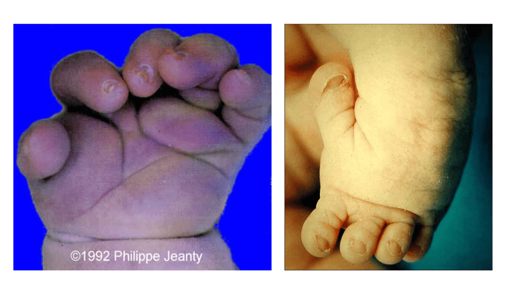
The characteristic hand deformity is the hitchhiker’s thumb, in which the thumb deviates away from the palm because of abnormal development of the first metacarpal and associated joints.
Other hand findings may include:
- Abnormal positioning of the fingers.
- Restricted movement of the interphalangeal joints.
- Contractures and reduced grip function.
- Abnormal spacing or configuration of the metacarpals.
These abnormalities may interfere with fine motor tasks, grasping, and other functional activities.
3. Ear Abnormalities
Swelling of the external ears is a classic early manifestation. The pinnae may become swollen during the neonatal period and subsequently develop a thickened, irregular, or cauliflower-like appearance.
These changes are related to abnormalities of cartilage and may persist as characteristic external ear deformities.
4. Cleft Palate and Facial Features
Cleft palate may occur and can contribute to feeding difficulties during infancy. Craniofacial findings vary, and not every affected individual has a cleft palate.
A careful newborn examination should assess the palate, external ears, hands, feet, and overall body proportions.
5. Foot Deformities
Foot deformities are among the most important causes of functional impairment in diastrophic dysplasia. Common abnormalities include:
- Congenital talipes equinovarus (clubfoot).
- Rigid or complex foot deformities.
- Skewfoot.
- Abnormal alignment of the hindfoot and forefoot.
The deformities can be severe and resistant to conservative treatment. A plantigrade foot, meaning a foot that can rest flat on the ground in a functional position, is an important treatment goal.
6. Joint Contractures
Contractures may affect the hips, knees, elbows, and other joints. They can be present early in life and may become more functionally limiting as the child grows.
Joint contractures may cause difficulty with walking, standing, transfers, dressing, and other everyday activities. Management focuses on maintaining useful range of motion, improving positioning, and reducing secondary deformity.
7. Hip and Knee Abnormalities
Hip involvement may include restricted motion, contractures, and abnormal joint development. Knee deformities, particularly genu valgum, may affect lower-limb alignment and gait.
Genu valgum is characterized by inward angulation of the knees, with the knees tending to approach one another when standing. Its clinical significance depends on the degree of deformity, symptoms, progression, and effect on walking.
8. Spinal Abnormalities
Spinal manifestations are clinically important because some may cause neurological complications.
Cervical spine: Cervical kyphosis may occur during early development. Atlantoaxial instability is a potentially serious complication because excessive movement between the first and second cervical vertebrae can threaten the spinal cord.
Thoracic and lumbar spine: Scoliosis and kyphoscoliosis may develop or progress with growth. Significant spinal deformity can affect posture, balance, mobility, and, in severe cases, respiratory function.
New weakness, changes in gait, loss of coordination, altered sensation, or other neurological symptoms require prompt assessment for possible spinal cord involvement.
Radiographic Findings
Radiographs are important for characterizing skeletal abnormalities, evaluating deformity, and planning orthopedic treatment. Findings should be interpreted alongside the clinical examination and, when appropriate, molecular genetic testing.
Common X-Ray Findings
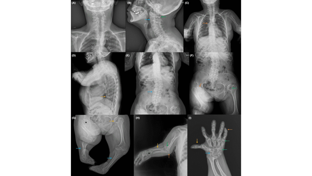
Typical skeletal imaging features may include:
- Generalized epiphyseal hypoplasia, with small or underdeveloped epiphyses.
- Epiphyseal flattening and irregularity.
- Metaphyseal widening or abnormal metaphyseal contour.
- Shortened long bones and abnormal limb proportions.
- Genu valgum and other lower-limb alignment abnormalities.
- Scoliosis or kyphoscoliosis.
- Abnormal configuration of the first metacarpal and widening of the first intermetacarpal space.
- Characteristic abnormalities of the hands and feet.
The exact findings depend on the patient’s age, the bones examined, and the severity of the disorder.
Spine Imaging
Spinal imaging is selected according to clinical findings and the suspected complication. Radiographs may demonstrate cervical kyphosis or scoliosis, while MRI is particularly valuable when there is concern about spinal cord compression or other neural structures.
When atlantoaxial instability is suspected, evaluation should be directed by a specialist familiar with skeletal dysplasia. Imaging positions and any assessment of cervical motion must be chosen carefully to avoid unnecessary risk in patients with possible instability.
Diagnosis of Diastrophic Dysplasia
Diagnosis is based on clinical findings, skeletal imaging, and molecular genetic testing.
1. Clinical Examination
Assessment begins with a detailed medical and family history and a physical examination. Features that may raise suspicion include:
- Disproportionate short stature.
- Hitchhiker’s thumbs.
- Characteristic external ear swelling or deformity.
- Congenital clubfoot or skewfoot.
- Multiple joint contractures.
- Cleft palate.
- Spinal curvature or abnormal cervical posture.
The examination should also document joint range of motion, limb alignment, gait where developmentally appropriate, and any neurological abnormalities.
2. Radiographic Assessment
Skeletal radiographs help identify characteristic abnormalities of the epiphyses, metaphyses, hands, feet, and spine. Additional imaging is guided by specific clinical concerns.
3. Molecular Genetic Testing
Identification of pathogenic variants in SLC26A2 can confirm the diagnosis. Testing may involve targeted analysis of the gene or a broader skeletal dysplasia panel, depending on the clinical presentation and availability of testing.
Genetic confirmation is particularly useful when clinical findings overlap with other skeletal dysplasias. It also supports family counseling and assessment of recurrence risk.
Differential Diagnosis
Several skeletal disorders may present with short stature, limb deformities, or abnormal cartilage development. Important considerations include:
Achondroplasia
Achondroplasia is commonly associated with disproportionate short stature and rhizomelic limb shortening, but it is caused by pathogenic variants in FGFR3 and has a different clinical and radiographic pattern.
Atelosteogenesis Type 2
Atelosteogenesis type 2 is another SLC26A2-related skeletal dysplasia. It is generally more severe and often presents with substantial skeletal abnormalities and life-threatening complications. Molecular and clinical assessment helps distinguish it from diastrophic dysplasia.
Spondyloepiphyseal Dysplasia
Spondyloepiphyseal dysplasias primarily affect the spine and epiphyses, although their presentation and associated features vary by subtype. The pattern of limb deformity, hand findings, spinal involvement, and genetic results helps establish the correct diagnosis.
Multiple Epiphyseal Dysplasia
Multiple epiphyseal dysplasia commonly causes joint pain, abnormal epiphyseal development, and early degenerative joint disease. The characteristic congenital hand and foot deformities of diastrophic dysplasia may help distinguish the two conditions.
A clinical geneticist or skeletal dysplasia specialist can help determine the most appropriate differential diagnosis and testing strategy.
Treatment and Management
There is no treatment that corrects the underlying genetic cause of diastrophic dysplasia. Management is directed toward improving function, preventing secondary complications, treating deformities, and maintaining mobility throughout growth.
Care is individualized and may involve pediatric orthopedists, physiotherapists, clinical geneticists, spinal specialists, rehabilitation professionals, and other clinicians as needed.
1. Physical Therapy and Rehabilitation
Physical therapy is an important component of early management. Goals include maintaining joint mobility, improving functional strength, supporting motor development, and optimizing posture and movement.
Therapy may include:
- Gentle range-of-motion exercises.
- Stretching for selected joint contractures.
- Functional strengthening and mobility training.
- Gait assessment and rehabilitation.
- Positioning strategies and appropriate assistive devices.
Exercises should be tailored to the individual’s skeletal abnormalities and should avoid forceful manipulation of potentially unstable joints, particularly the cervical spine.
2. Management of Joint Contractures
Early assessment of contractures can help guide conservative treatment and determine whether surgery is necessary.
Physical therapy, positioning, and selected orthotic interventions may improve function in some patients. However, established or severe contractures may require surgical release.
Hip, knee, and elbow procedures are selected according to the location and severity of the contracture, functional limitations, and the patient’s overall skeletal anatomy.
3. Treatment of Foot Deformities
Rigid clubfoot and other complex foot deformities can be difficult to manage. The objective is to obtain a stable, plantigrade, comfortable foot that facilitates standing, walking, and footwear use.
Treatment may include carefully selected casting, orthoses, and surgical correction. Severe rigid deformities may require extensive soft-tissue releases or other reconstructive procedures.
The specific approach should be determined by a pediatric orthopedic surgeon experienced in complex congenital foot deformities. Recurrence, stiffness, skin problems, and altered growth must be considered during long-term follow-up.
4. Management of Cervical Spine Abnormalities
Cervical spine assessment is essential because atlantoaxial instability and cervical deformity may threaten neurological function.
Patients with suspected instability require specialist evaluation. Neurological symptoms associated with instability or spinal cord compression may necessitate stabilization, including occipitocervical fusion in appropriately selected cases.
Cervical kyphosis may improve spontaneously in some children, but this should not be assumed in every patient. Progressive deformity, persistent compression, or neurological compromise may require further intervention, potentially including corrective fusion.
Management decisions depend on spinal alignment, stability, symptoms, growth, and imaging findings.
5. Treatment of Scoliosis and Kyphoscoliosis
Spinal curvature should be monitored during growth. Treatment depends on the magnitude and progression of the curve, skeletal maturity, symptoms, and any associated neurological or respiratory problems.
Observation and selected nonoperative measures may be appropriate in some patients. Progressive or severe deformity may require spinal fusion, often using a posterior approach when indicated.
Surgical planning is complex in skeletal dysplasia and requires careful evaluation of spinal anatomy, bone quality, neurological risk, and the potential effect of surgery on future growth and function.
6. Management of Hip and Knee Deformities
Hip and knee deformities are evaluated according to symptoms, functional limitation, joint range of motion, and alignment.
Treatment may include rehabilitation, orthoses, or corrective surgery for selected patients. The decision to operate should consider the child’s growth, the nature of the deformity, gait mechanics, and the likelihood of improving function.
7. Management of Cleft Palate and Other Associated Problems
When cleft palate is present, care may involve a cleft team, including surgical, feeding, speech, and dental specialists. Feeding support may be particularly important during infancy.
Hearing, breathing, sleep, and developmental concerns should be assessed when indicated by the patient’s symptoms and clinical findings.
8. Long-Term Follow-Up
Diastrophic dysplasia requires continuing care because deformities and functional limitations may change with growth. Follow-up should assess:
- Height and skeletal growth.
- Joint range of motion and contractures.
- Foot alignment and walking ability.
- Hip and knee deformities.
- Spinal curvature and cervical stability.
- Neurological symptoms.
- Pain, participation, and activities of daily living.
A coordinated plan helps clinicians identify complications early and adjust treatment as the child develops.
Prognosis
The prognosis of diastrophic dysplasia varies according to the severity of skeletal involvement and the presence of complications. Many affected individuals survive into adulthood, but short stature, joint deformities, restricted mobility, and spinal abnormalities may lead to substantial lifelong functional challenges.
Early orthopedic assessment, appropriate rehabilitation, and timely treatment of progressive deformities can improve mobility and independence. Cervical spine complications deserve particular attention because spinal cord compression can result in serious neurological injury.
Long-term outcomes depend on the individual’s anatomy, the severity of contractures and deformities, access to specialized care, and the success of interventions. Regular monitoring and individualized treatment are therefore essential.
Key Clinical Points
- Diastrophic dysplasia is an autosomal recessive skeletal dysplasia caused by pathogenic variants in SLC26A2.
- Defective sulfate transport disrupts cartilage proteoglycan sulfation and normal skeletal development.
- Typical findings include disproportionate short stature, hitchhiker’s thumbs, characteristic ear deformities, clubfoot, and joint contractures.
- Radiographs may show epiphyseal hypoplasia and flattening, metaphyseal abnormalities, genu valgum, and spinal deformities.
- Diagnosis combines clinical assessment, skeletal imaging, and molecular genetic testing.
- Physical therapy and individualized orthopedic management are central to treatment.
- Atlantoaxial instability and progressive spinal deformity require careful assessment because of the risk of neurological complications.
- Long-term multidisciplinary follow-up supports mobility, function, and quality of life.
Frequently Asked Questions (FAQ)
What causes diastrophic dysplasia?
Diastrophic dysplasia is caused by pathogenic variants in the SLC26A2 gene. These variants impair sulfate transport, which is necessary for normal cartilage proteoglycan formation and skeletal development.
Is diastrophic dysplasia inherited?
Yes. It is inherited in an autosomal recessive pattern. An affected individual typically inherits one pathogenic variant from each parent.
What are the characteristic features of diastrophic dysplasia?
Characteristic findings include disproportionate short stature, hitchhiker’s thumbs, cauliflower-like external ear deformities, congenital clubfoot, joint contractures, and possible cleft palate. Spinal abnormalities may include cervical kyphosis and scoliosis.
How is diastrophic dysplasia diagnosed?
Diagnosis is based on clinical findings, skeletal radiographs, and molecular genetic testing to identify pathogenic variants in SLC26A2.
What are the radiographic findings in diastrophic dysplasia?
Radiographs may demonstrate epiphyseal hypoplasia and flattening, metaphyseal widening, shortened long bones, genu valgum, scoliosis, and characteristic hand and foot abnormalities.
Can diastrophic dysplasia be cured?
There is currently no established treatment that corrects the underlying genetic defect. Management focuses on treating deformities, maintaining joint mobility, preventing complications, and improving function.
Does diastrophic dysplasia require surgery?
Some patients require orthopedic surgery for severe contractures, rigid foot deformities, progressive spinal curvature, or cervical instability associated with neurological risk. The need for surgery depends on the individual’s symptoms, anatomy, and functional limitations.
What is the significance of atlantoaxial instability in diastrophic dysplasia?
Atlantoaxial instability involves excessive movement between the first and second cervical vertebrae. In severe cases, it can cause spinal cord compression and neurological injury, making specialist assessment important when instability is suspected or symptoms develop.
What is the life expectancy of a person with diastrophic dysplasia?
Life expectancy varies with disease severity and associated complications. Many affected individuals live into adulthood, although significant orthopedic and mobility challenges may persist. Individual prognosis should be discussed with the treating clinical team.
Is genetic counseling recommended for diastrophic dysplasia?
Yes. Genetic counseling helps explain autosomal recessive inheritance, carrier testing, recurrence risks for future pregnancies, and reproductive options for affected individuals and their families.
References
- National Center for Biotechnology Information (NCBI), GeneReviews and related resources on SLC26A2-related skeletal dysplasias. https://www.ncbi.nlm.nih.gov/books/
- National Library of Medicine, PubMed: research literature on diastrophic dysplasia, SLC26A2, and skeletal dysplasia. https://pubmed.ncbi.nlm.nih.gov/?term=diastrophic+dysplasia
- National Library of Medicine, PubMed: research on SLC26A2-related disorders and sulfate transporter function. https://pubmed.ncbi.nlm.nih.gov/?term=SLC26A2+skeletal+dysplasia
- MedlinePlus Genetics, Diastrophic dysplasia. https://medlineplus.gov/genetics/