Hyperparathyroidism is an endocrine disorder characterized by excessive secretion of parathyroid hormone (PTH), a hormone that regulates calcium and phosphate levels in the blood. The condition can affect the kidneys, bones, gastrointestinal system, and muscles. In some patients, hyperparathyroidism causes few noticeable symptoms and is discovered incidentally during routine blood tests. In others, it may lead to kidney stones, reduced bone density, muscle weakness, and characteristic skeletal abnormalities.
Understanding the different types of hyperparathyroidism, their clinical manifestations, and their radiographic findings is important for medical professionals, particularly those involved in endocrinology, orthopedics, and metabolic bone disease.
What Is Hyperparathyroidism?
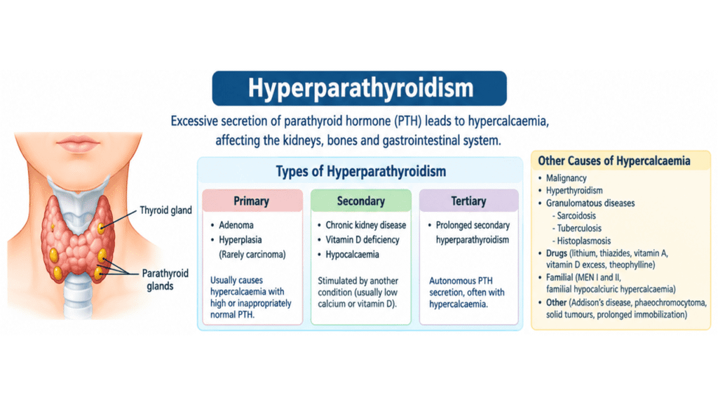
The parathyroid glands are four small endocrine glands located behind the thyroid gland. They produce PTH, which helps maintain calcium homeostasis by regulating calcium reabsorption in the kidneys, activating vitamin D to increase intestinal calcium absorption, and stimulating bone remodeling.
Hyperparathyroidism develops when the parathyroid glands release excessive amounts of PTH. The resulting effects depend on the underlying cause and duration of the disorder.
Types of Hyperparathyroidism
1. Primary hyperparathyroidism
Primary hyperparathyroidism results from abnormal activity of one or more parathyroid glands. The most common cause is a solitary parathyroid adenoma, while less common causes include multiglandular hyperplasia and, rarely, parathyroid carcinoma. It frequently presents with elevated serum calcium and an elevated or inappropriately normal PTH level.
2. Secondary hyperparathyroidism
Secondary hyperparathyroidism develops when another condition persistently stimulates PTH secretion, usually because of low calcium availability or impaired vitamin D metabolism. Chronic kidney disease is a major cause. Reduced phosphate excretion, impaired activation of vitamin D, and disturbances in calcium balance contribute to increased PTH secretion.
3. Tertiary hyperparathyroidism
Tertiary hyperparathyroidism occurs when prolonged secondary hyperparathyroidism leads to autonomous parathyroid hormone secretion. It is most commonly associated with longstanding chronic kidney disease, particularly after prolonged secondary hyperparathyroidism. Unlike uncomplicated secondary hyperparathyroidism, tertiary disease often produces hypercalcemia.
Pathophysiology of Hyperparathyroidism
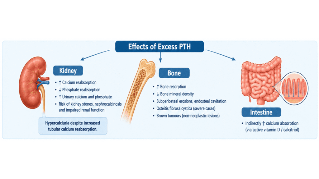
Excessive PTH affects calcium and phosphate metabolism through several mechanisms:
- Kidneys: PTH increases calcium reabsorption in the renal tubules while reducing phosphate reabsorption, resulting in increased urinary phosphate excretion.
- Intestines: PTH indirectly increases intestinal calcium absorption by stimulating the renal production of active vitamin D, or calcitriol.
- Bones: PTH increases bone remodeling and, when its effects are excessive or sustained, promotes bone resorption and the release of calcium and phosphate from the skeleton.
In primary hyperparathyroidism, increased serum calcium can lead to hypercalciuria despite enhanced tubular calcium reabsorption. This increases the risk of kidney stones and, in some patients, nephrocalcinosis. Persistent calcium disturbances may also impair renal function.
In the skeleton, excessive PTH activity can cause reduced bone mineral density, cortical bone loss, and increased fracture risk. Severe, longstanding disease may produce osteitis fibrosa cystica, a condition characterized by marked bone resorption, fibrous tissue replacement, and cyst-like lesions.
What Is Osteitis Fibrosa Cystica?
Osteitis fibrosa cystica is a severe skeletal manifestation of hyperparathyroidism. Increased osteoclastic bone resorption produces areas of cortical erosion and bone destruction. The marrow spaces may be replaced by vascular fibrous tissue, and hemorrhage with a giant-cell reaction can produce brownish lesions known as brown tumors.
Despite their name, brown tumors are not true neoplasms. They represent localized, non-neoplastic bone lesions associated with excessive PTH activity. They may resemble primary bone tumors, metastatic lesions, or other cystic bone abnormalities on imaging, making biochemical evaluation essential for diagnosis.
Causes of Hypercalcemia
Hyperparathyroidism is an important cause of hypercalcemia, but elevated serum calcium can result from several other disorders. Identifying the underlying cause is essential because treatment differs substantially between conditions.
Major causes include:
- Increased bone resorption: Primary or tertiary hyperparathyroidism, malignancy, and prolonged immobilization.
- Malignancy: Certain cancers cause hypercalcemia through PTH-related peptide production, bone destruction, or other mechanisms.
- Granulomatous diseases: Sarcoidosis, tuberculosis, and histoplasmosis can increase active vitamin D production in affected tissues.
- Medications and supplements: Lithium, thiazide diuretics, excessive vitamin D, vitamin A toxicity, and, in some circumstances, theophylline toxicity.
- Familial conditions: Familial hypocalciuric hypercalcemia and hereditary endocrine syndromes, including multiple endocrine neoplasia (MEN).
- Other endocrine disorders: Addison disease and hyperthyroidism.
- Other causes: Certain solid tumors and disorders associated with altered calcium metabolism.
Familial hypocalciuric hypercalcemia is particularly important in the differential diagnosis because it can resemble primary hyperparathyroidism but generally causes lifelong, mild hypercalcemia with relatively low urinary calcium excretion. Unnecessary parathyroid surgery should be avoided when this condition is correctly identified.
Primary Hyperparathyroidism
Primary hyperparathyroidism is the most common form of hyperparathyroidism encountered in general endocrine practice. It is frequently caused by a single benign parathyroid adenoma, although hyperplasia affecting multiple glands can also occur.
The condition is often diagnosed in middle-aged and older adults and is more common in women. Many patients have no obvious symptoms, and the diagnosis is made after routine blood testing reveals elevated serum calcium.
Clinical Features and Symptoms
Symptoms vary according to the severity and duration of hypercalcemia and the degree of skeletal involvement.
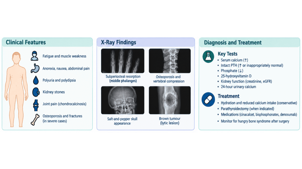
Common clinical features include:
- Fatigue and generalized weakness.
- Loss of appetite, nausea, and abdominal discomfort.
- Constipation and, in some patients, other gastrointestinal symptoms.
- Increased thirst and frequent urination.
- Kidney stones and nephrocalcinosis.
- Reduced bone mineral density and osteoporosis.
- Bone pain and an increased risk of fractures in advanced disease.
- Depression, difficulty concentrating, or other nonspecific neurocognitive complaints.
- Chondrocalcinosis, which may cause joint pain or acute inflammatory joint symptoms.
Severe skeletal manifestations, such as osteitis fibrosa cystica and brown tumors, are uncommon in patients diagnosed through routine biochemical screening. Nevertheless, they remain important findings in advanced or longstanding disease.
Renal Complications
The kidneys are particularly vulnerable to disturbances in calcium metabolism. Increased urinary calcium excretion can promote the formation of calcium-containing kidney stones. Calcium deposition within renal tissue, known as nephrocalcinosis, may occur in more advanced cases.
Patients may also experience polyuria and excessive thirst. Persistent renal complications can contribute to impaired kidney function, emphasizing the importance of assessing renal status during evaluation.
Skeletal Complications
Excessive PTH activity increases bone turnover and can reduce bone mineral density. Cortical bone, including the distal radius, is particularly important when assessing skeletal involvement in primary hyperparathyroidism.
Although severe bone disease is now less commonly encountered in many clinical settings, patients may develop osteoporosis, vertebral compression fractures, subperiosteal bone resorption, and brown tumors. Bone mineral density testing helps identify skeletal involvement and assess fracture risk.
X-Ray Findings of Hyperparathyroidism
Radiographic imaging can reveal characteristic skeletal changes in patients with longstanding hyperparathyroidism. However, normal X-rays do not exclude the diagnosis, particularly in patients with early or mild disease.
1. Subperiosteal Bone Resorption
Subperiosteal bone resorption is a classic radiographic finding of hyperparathyroidism. It is particularly noticeable along the radial aspects of the middle phalanges of the fingers. This results from excessive bone resorption beneath the periosteum and is an important clue when evaluating suspected metabolic bone disease.
2. Osteoporosis and Cortical Bone Loss
Generalized bone loss may occur, with cortical thinning and reduced bone density. Vertebral compression fractures can develop in patients with significant skeletal involvement. Dual-energy X-ray absorptiometry (DXA) is used to measure bone mineral density and evaluate osteoporosis.
3. Brown Tumors
Brown tumors appear as well-defined or expansile osteolytic lesions that may resemble bone cysts, giant-cell tumors, or metastatic disease. They are caused by localized bone remodeling, hemorrhage, and fibrous tissue proliferation associated with excessive PTH activity. Biochemical testing is essential to distinguish these lesions from other bone disorders.
4. Other Radiographic Findings
Additional findings may include:
- Endosteal bone resorption.
- Subperiosteal cortical erosions.
- Acro-osteolysis in advanced disease.
- Salt-and-pepper appearance of the skull due to heterogeneous bone resorption.
- Renal calculi and nephrocalcinosis on appropriate imaging.
- Chondrocalcinosis, particularly involving articular cartilage and fibrocartilage.
These findings are not present in every patient. Modern laboratory testing and DXA frequently detect disease before severe skeletal abnormalities become apparent.
Diagnosis of Hyperparathyroidism
The diagnosis is based on clinical assessment, biochemical testing, and evaluation for complications. Imaging may help assess the effects of the disease or locate abnormal parathyroid tissue before surgery, but it does not replace biochemical confirmation.
Blood Tests
Important laboratory investigations include:
- Serum calcium: Elevated in most cases of primary hyperparathyroidism.
- Intact PTH: Elevated or inappropriately normal in primary hyperparathyroidism despite hypercalcemia.
- Serum phosphate: Often reduced in primary hyperparathyroidism because PTH increases renal phosphate excretion.
- 25-hydroxyvitamin D: Helps identify vitamin D deficiency and guide management.
- Serum creatinine and estimated glomerular filtration rate (eGFR): Assess kidney function.
- Alkaline phosphatase: May be elevated when bone turnover is substantially increased.
- 24-hour urinary calcium: Helps assess urinary calcium excretion and distinguish selected alternative diagnoses, including familial hypocalciuric hypercalcemia.
If hypercalcemia is present with a suppressed PTH level, other causes, such as malignancy, vitamin D-related disorders, and granulomatous diseases, should be considered.
Imaging and Bone Assessment
Additional investigations may include DXA scans of the lumbar spine, hip, and distal one-third of the radius, vertebral fracture assessment, and renal imaging to identify kidney stones or nephrocalcinosis.
Parathyroid ultrasound, sestamibi scintigraphy, or other localization studies may be used when surgery is planned. These investigations help surgeons locate abnormal glands; they are not diagnostic tests for hyperparathyroidism by themselves.
Treatment of Hyperparathyroidism
Treatment depends on the underlying cause, the severity of biochemical abnormalities, and the presence of kidney or skeletal complications.
Conservative Management
Selected patients with mild primary hyperparathyroidism who do not meet surgical criteria may be monitored regularly. Follow-up generally includes serum calcium, kidney function, vitamin D assessment, and periodic bone mineral density measurements.
Patients should maintain adequate hydration, remain physically active when appropriate, and avoid dehydration. Dietary calcium should generally follow recommended nutritional requirements rather than being severely restricted without medical advice. Vitamin D deficiency should be managed carefully under clinical supervision.
Parathyroidectomy
Parathyroidectomy is the definitive treatment for primary hyperparathyroidism when surgery is indicated. The procedure removes the abnormal parathyroid gland or glands and can normalize calcium and PTH levels.
Surgery is generally recommended for symptomatic disease and may be indicated in otherwise asymptomatic patients who meet established criteria, including significant hypercalcemia, osteoporosis or vertebral fracture, kidney stones, impaired renal function, hypercalciuria meeting guideline thresholds, or age below 50 years. Individual decisions should follow current clinical guidelines and specialist assessment.
Medical Treatment
When surgery is unsuitable or deferred, medication may be considered according to the patient’s clinical needs.
- Cinacalcet: Reduces PTH secretion and can lower serum calcium.
- Bisphosphonates: May improve bone mineral density in selected patients.
- Denosumab: May be considered in selected cases to improve bone density.
- Vitamin D supplementation: May be appropriate when deficiency is present, with careful monitoring of calcium levels.
These treatments have different effects and are not interchangeable. Medication choice depends on whether the primary treatment goal is controlling hypercalcemia, improving bone density, or managing the underlying cause.
Hungry Bone Syndrome After Surgery
Following parathyroidectomy, some patients develop significant hypocalcemia because previously high-turnover bone rapidly takes up calcium, phosphate, and magnesium during remineralization. This complication is known as hungry bone syndrome.
Patients with severe skeletal disease or markedly elevated bone turnover may be at increased risk. Management may require close biochemical monitoring and calcium, active vitamin D, and sometimes magnesium supplementation. Prompt recognition is important to prevent complications associated with low serum calcium.
Secondary and Tertiary Hyperparathyroidism: Treatment Considerations
Secondary hyperparathyroidism is managed by treating the underlying cause. In chronic kidney disease, management may involve correcting vitamin D deficiency, addressing phosphate abnormalities, reviewing calcium balance, and using selected medications under specialist supervision.
Tertiary hyperparathyroidism may require more intensive medical treatment or parathyroidectomy when autonomous PTH secretion causes persistent hypercalcemia or other complications. Treatment decisions are particularly important in patients with advanced kidney disease or a history of kidney transplantation.
Frequently Asked Questions (FAQ)
1. What is the most common cause of primary hyperparathyroidism?
A solitary benign parathyroid adenoma is the most common cause. Multiglandular hyperplasia is another important cause, while parathyroid carcinoma is rare.
2. What are the classic symptoms of hyperparathyroidism?
Common symptoms include fatigue, muscle weakness, constipation, nausea, increased thirst, frequent urination, and bone or joint discomfort. Some patients remain asymptomatic and are diagnosed through routine blood tests.
3. What is the difference between primary, secondary, and tertiary hyperparathyroidism?
Primary hyperparathyroidism originates from abnormal activity of the parathyroid glands. Secondary hyperparathyroidism develops in response to another condition, commonly chronic kidney disease or vitamin D deficiency. Tertiary hyperparathyroidism occurs when prolonged secondary stimulation leads to autonomous PTH secretion.
4. What is the classic X-ray finding of hyperparathyroidism?
Subperiosteal bone resorption along the radial aspects of the middle phalanges is a classic finding. Other abnormalities include cortical thinning, osteoporosis, brown tumors, and a salt-and-pepper appearance of the skull.
5. What are brown tumors in hyperparathyroidism?
Brown tumors are non-neoplastic bone lesions caused by excessive bone remodeling associated with elevated PTH. They may contain fibrous tissue, hemorrhage, and multinucleated giant cells. Their appearance can mimic other bone lesions.
6. Can hyperparathyroidism cause kidney stones?
Yes. Increased urinary calcium excretion can promote calcium-containing kidney stones. Persistent calcium deposition in the kidneys may also cause nephrocalcinosis and, in some cases, impaired renal function.
7. Which blood tests are used to diagnose hyperparathyroidism?
The main tests include serum calcium, intact PTH, phosphate, 25-hydroxyvitamin D, and kidney function measurements. A 24-hour urinary calcium test may help distinguish alternative causes of hypercalcemia.
8. Is surgery always necessary for primary hyperparathyroidism?
No. Parathyroidectomy is the definitive treatment when indicated, but selected patients with mild disease may undergo structured monitoring. Surgery is generally recommended for symptomatic disease and for patients meeting established biochemical, renal, or skeletal criteria.
9. What is hungry bone syndrome?
Hungry bone syndrome is a postoperative complication in which bone rapidly absorbs calcium and other minerals after parathyroidectomy, causing prolonged or severe hypocalcemia. It requires prompt monitoring and appropriate treatment.
10. Can hyperparathyroidism cause osteoporosis?
Yes. Excessive PTH activity can increase bone turnover and reduce bone mineral density, particularly at cortical bone sites. Severe disease can also cause fractures, subperiosteal bone resorption, and osteitis fibrosa cystica.
References
- Bilezikian JP, Khan AA, Silverberg SJ, et al. Evaluation and Management of Primary Hyperparathyroidism: Summary Statement and Guidelines from the Fifth International Workshop. Journal of Bone and Mineral Research. 2022;37(11):2293–2314. Read on PubMed.
- Kochman M. Primary hyperparathyroidism: clinical manifestations, diagnosis and evaluation according to the Fifth International Workshop guidelines. Reumatologia. 2023. Read on PubMed.
- National Institute of Diabetes and Digestive and Kidney Diseases (NIDDK). Primary Hyperparathyroidism. Read the clinical overview.
- Management of Primary Hyperparathyroidism. Journal of Bone and Mineral Research. 2022;37(11):2391–2403. Read on PubMed.
- Orthofixar. Orthopedic and clinical examination educational resources.