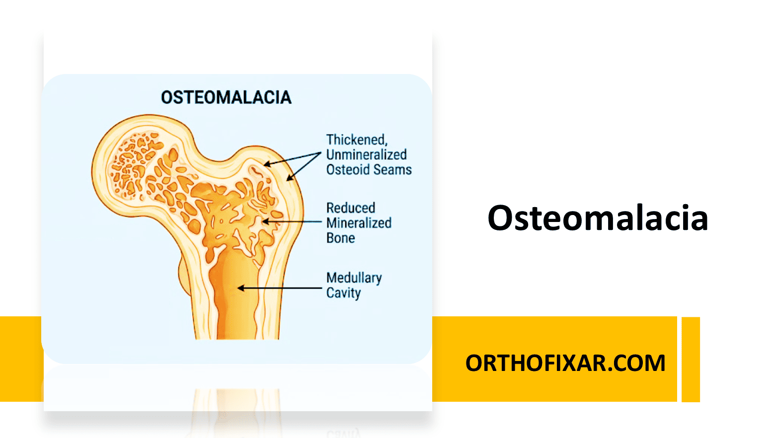
What Is Osteomalacia?
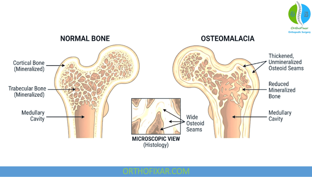
Osteomalacia is a metabolic bone disorder in adults in which bone does not mineralize properly, so it becomes soft. The most common cause is vitamin D deficiency. Other causes include malabsorption, chronic kidney disease and low phosphate. Because the condition develops slowly, it is often missed or mistaken for other musculoskeletal problems (Bhan et al., PubMed).
Common Symptoms of Osteomalacia
Osteomalacia can have an insidious course. Early symptoms are vague and non-specific, which delays diagnosis. Typical complaints include:
- Widespread bone pain, often in the hips, legs, ribs or lower back
- Muscle weakness, usually in the proximal muscles around the hips and shoulders
- A “waddling” gait, caused by that proximal weakness
- Unexplained hip or long-bone pain, which may warn of a stress fracture
- A higher fracture risk throughout the skeleton
Persistent, unexplained bone pain or weakness should be assessed by a doctor.
Looser Zones: The Classic Sign on X-Ray
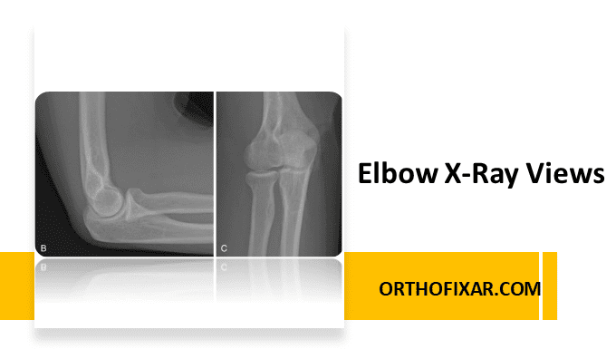
The classic radiological sign is the Looser zone, also called a pseudofracture or Milkman line. It is a thin, transverse band of rarefaction in otherwise normal-looking bone. Looser zones are seen especially in the:
- Pubic rami
- Medial proximal femur
- Axillary (outer) edge of the scapula
They are incomplete stress fractures. They heal with callus that lacks calcium, so the bone never fully strengthens.
Vertebral fractures in osteomalacia can look biconcave or wedge-shaped. These changes are hard to tell apart from osteoporotic fractures on imaging, so blood tests and clinical context matter. Signs of secondary hyperparathyroidism may also be present. A raised alkaline phosphatase, low calcium or phosphate, and low serum 25-hydroxyvitamin D (25-OHD) all support the diagnosis (Holick, 2011, PubMed).
Can Osteomalacia Be Prevented?
In high-risk groups, osteomalacia is generally preventable through diet changes or vitamin D supplements. UK government guidance recommends 400 IU of vitamin D daily for high-risk groups such as pregnant women (Gov.uk, 2012).
Patients with malabsorption may need larger doses. Their 25-OHD levels should be checked to confirm adequate replacement.
Calcium and Vitamin D Together
Most vitamin D products are combined with calcium. This can help, because calcium deficiency may worsen the effects of vitamin D deficiency. However, calcium deficiency that needs treatment in its own right is uncommon. Patients with malabsorption or inflammatory bowel disease are at particular risk.
Some people cannot tolerate combined supplements because of upper gastrointestinal side effects. The calcium component is usually the cause, and vitamin D-only supplements can overcome the problem.
Osteomalacia Treatment: Vitamin D Loading Doses
After diagnosis, long-term vitamin D supplementation is generally required. Treatment starts with a loading dose of roughly 300,000–600,000 IU to replenish body stores. The risk of hypercalcaemia is low, and the dose is given in one of two ways:
- Depot injections (for example, ergocalciferol) given twice, four weeks apart
- High-strength oral vitamin D (10,000 IU or 50,000 IU doses) over an equivalent period
More recently, the bioavailability of vitamin D after intramuscular depot injection has been questioned. High-strength oral vitamin D is now generally favoured for loading. Treatment should always be supervised by a healthcare professional, with follow-up blood tests to confirm vitamin D levels are adequate.
Frequently Asked Questions (FAQ)
What is the main cause of osteomalacia?
Vitamin D deficiency is the most common cause. It can result from low sun exposure, poor diet, or malabsorption. Kidney disease and phosphate problems are other causes.
What are the first signs of osteomalacia?
Early signs are non-specific. They include diffuse bone pain, proximal muscle weakness and, sometimes, a waddling walk.
What is a Looser zone?
A Looser zone is a thin band of reduced bone density running across the bone on X-ray. It is an incomplete stress fracture that healed with calcium-poor callus, and it is the classic lesion of osteomalacia.
How is osteomalacia different from osteoporosis?
In osteomalacia, bone is poorly mineralized. In osteoporosis, bone mass is reduced but mineralization is normal. Vertebral fractures can look similar on imaging, so blood tests (25-OHD, calcium, phosphate, alkaline phosphatase) help distinguish them.
Can osteomalacia be prevented?
Usually, yes. In high-risk groups, dietary changes and vitamin D supplements are effective. The UK recommendation for groups such as pregnant women is 400 IU daily.
Do I need calcium as well as vitamin D?
Combined products are common and can help. If calcium upsets your stomach, vitamin D-only supplements are an option.
How is osteomalacia treated?
Treatment is a vitamin D loading dose (about 300,000–600,000 IU, now usually as high-strength oral doses), followed by long-term maintenance.
Is a vitamin D loading dose safe?
The risk of hypercalcaemia is low, but loading doses should only be taken under medical supervision.
References
- Holick MF et al. Evaluation, treatment, and prevention of vitamin D deficiency: an Endocrine Society clinical practice guideline. J Clin Endocrinol Metab. 2011. PubMed 21646368
- Holick MF. Vitamin D deficiency. N Engl J Med. 2007. PubMed 17634462
- Bhan A, Rao AD, Rao DS. Osteomalacia as a result of vitamin D deficiency. Endocrinol Metab Clin North Am. 2010. PubMed 20511055
- Gov.uk (2012), vitamin D recommendation for high-risk groups, as cited in your source text.