The Kendall Test, also known as the Rectus Femoris Contracture Test, is a clinical examination used to assess shortening or contracture of the rectus femoris muscle. Because the rectus femoris crosses both the hip and knee joints, tightness typically affects both hip extension and knee flexion.
The rectus femoris is one of the quadriceps group, as it is the only muscle that crosses both the hip and knee joints. Originating from the anterior inferior iliac spine and the superior rim of the acetabulum, it inserts into the patella via the quadriceps tendon and ultimately onto the tibial tuberosity through the patellar ligament. This biarticular nature means that contracture of the rectus femoris can significantly impact both hip extension and knee flexion mobility.
Clinical Significance: Rectus femoris contracture can contribute to anterior pelvic tilt, lumbar lordosis, knee pain, and altered gait patterns. Early identification through the Kendall Test allows for targeted intervention and improved functional outcomes.
See Also: Rectus Femoris Muscle Anatomy
How to Perform the Kendall Test?
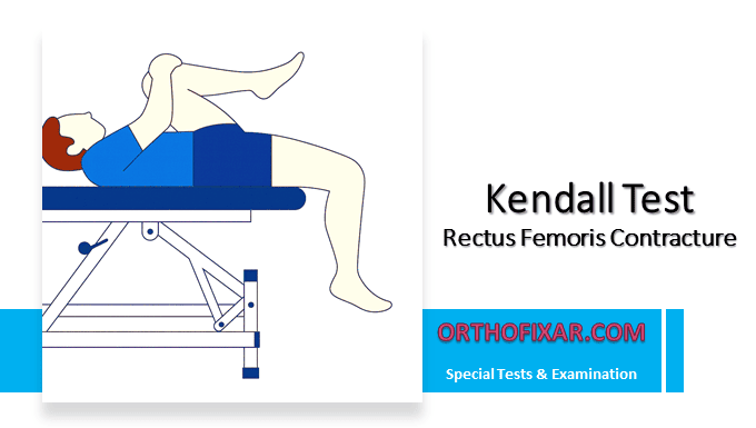
- The patient lies supine on the examining table with the knees hanging over the edge. This position allows the knee to move freely into flexion or extension during the test.
- Ask the patient to flex one knee toward the chest and hold it firmly.
- Observe the opposite leg, which becomes the test limb.
- Evaluate the angle of the test knee:
- A normal response is for the test knee to remain at 90° of flexion when the opposite hip is fully flexed.
- If the knee begins to extend slightly, this suggests a rectus femoris contracture.
- The examiner may passively flex the test knee to confirm whether it naturally returns to 90°.
- Always palpate the rectus femoris during the maneuver to detect true muscle tightness.
See Also: Thomas Test

What does a Positive Kendall Test Mean?
The Kendall Test is positive when the tested knee extends instead of holding at 90° suggesting tightness or contracture of the rectus femoris.
| Finding | Interpretation | Clinical Implication |
|---|---|---|
| Negative Test | Test knee maintains 90° flexion | Normal rectus femoris length and flexibility |
| Positive Test | Test knee extends beyond 90° | Suggests rectus femoris contracture or tightness |
| Palpable Muscle Tightness | Tension felt in muscle belly | Muscular origin of restriction; muscle stretch end feel |
| No Palpable Tightness | Restriction without muscle tension | Likely capsular restriction; capsular end feel |
Differentiating Muscle Tightness vs. Joint Restriction
Muscular Restriction Characteristics:
When palpating during the test, muscular restriction presents with palpable tightness along the muscle belly and tendon. The end feel is typically described as a firm, elastic resistance that has some give to it. Patients may report a stretching sensation along the anterior thigh. Muscular restrictions generally respond well to stretching, soft tissue mobilization, and progressive strengthening through full range of motion.
Capsular Restriction Characteristics:
In contrast, capsular restrictions occur without significant palpable muscle tightness. The end feel is characteristically firm and has less give compared to muscular restrictions, often described as a harder, more abrupt stop to movement. Capsular patterns may be associated with previous injury, prolonged immobilization, or degenerative joint changes. Treatment typically requires joint mobilization techniques, prolonged low-load stretching, and addressing underlying joint pathology.
In summary:
- Muscle tightness:
- Palpable increased tension
- A muscular end-feel (stretch-type)
- Joint or capsular restriction:
- No palpable muscle tightness
- A firmer, capsular end-feel.
Clinical Notes
- Both sides must be tested and compared for asymmetries.
- The Kendall Test is particularly useful for evaluating patients with:
- Anterior knee pain
- Hip flexor imbalance
- Lumbar spine compensations related to pelvic tilt
- Always assess the patient’s comfort and maintain proper stabilization to avoid compensatory movements.
References & More
- Orthopedic Physical Assessment by David J. Magee, 7th Edition.
- Murdock CJ, Mudreac A, Agyeman K. Anatomy, Abdomen and Pelvis, Rectus Femoris Muscle. [Updated 2023 Nov 13]. In: StatPearls [Internet]. Treasure Island (FL): StatPearls Publishing; 2025 Jan-. Available from: PubMed
