What Is Albers–Schönberg Disease?
Albers–Schönberg disease generally refers to autosomal dominant osteopetrosis type II (ADOII), an inherited skeletal disorder caused by impaired osteoclast function. Bone becomes unusually dense on radiographs, yet remains vulnerable to fractures because normal bone remodeling is disrupted. The condition belongs to the broader osteopetrosis family, also known as marble bone disease.
Although traditionally described as “adult” or “tarda” osteopetrosis, Albers–Schönberg disease can become apparent during childhood or adolescence. Its severity varies considerably, including among relatives with the same genetic variant. Some individuals are diagnosed incidentally, while others develop recurrent fractures, persistent pain, dental infection, or nerve compression.
Who Is Albers–Schönberg Disease Named After?
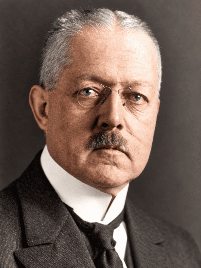
Albers–Schönberg disease is named after Heinrich Ernst Albers–Schönberg (1865–1921), a German physician and pioneer of radiology. In 1904, he described the characteristic X-ray findings of unusually dense bones in a young man with fractures. His report established the radiographic description of the condition historically known as marble bone disease.
What Causes Albers–Schönberg Disease?
CLCN7 Variants and Defective Bone Resorption
Albers–Schönberg disease is most commonly associated with a heterozygous pathogenic variant in the CLCN7 gene. This gene encodes ClC-7, a chloride–proton exchanger that supports the ionic conditions required for osteoclast-mediated bone resorption.
When ClC-7 function is disrupted, osteoclasts cannot remove bone effectively. Bone formation continues while resorption is impaired, allowing abnormal bone to accumulate. This produces osteosclerosis and can reduce the space available for marrow. Increased mineral content therefore coexists with abnormal skeletal architecture and fragility.
Inheritance and Genetic Counseling
An individual with a disease-causing autosomal dominant CLCN7 variant has a 50% chance of transmitting the variant in each pregnancy. Inheritance does not reliably predict severity: reduced penetrance means some carriers lack recognizable disease, and variable expressivity means affected relatives may have different complications. Genetic counseling helps families interpret these risks.
Types of Osteopetrosis: Adult Versus Infantile Disease
Osteopetrosis comprises several genetically distinct conditions. Classification by age alone is insufficient because clinical features overlap.
| Form | Typical presentation | Main clinical concerns |
|---|---|---|
| Severe autosomal recessive osteopetrosis | Infancy | Growth impairment, marrow failure, cranial nerve compression, and metabolic complications |
| Intermediate osteopetrosis | Childhood | Variable fractures, anemia, and nerve involvement; inheritance can be recessive or dominant |
| Autosomal dominant osteopetrosis type II | Often recognized in later childhood, adolescence, or adulthood | Fragility fractures, pain, jaw infection, and occasional neurological or hematological complications |
Historical estimates suggest a frequency of approximately 1 in 20,000 for autosomal dominant osteopetrosis and 1 in 250,000 births for autosomal recessive osteopetrosis. These figures vary by population and should not be treated as one universal incidence estimate.
In severe infantile disease, extensive skeletal sclerosis can restrict marrow space, causing anemia, thrombocytopenia, and sometimes leukopenia. Blood production outside the marrow can enlarge the liver and spleen. Growth failure, visual impairment, hearing loss, hydrocephalus, and hypocalcemia may also occur. Secondary hyperparathyroidism can accompany hypocalcemia; an elevated parathyroid hormone level is not a universal feature of every osteopetrosis subtype.
Albers–Schönberg Disease Symptoms and Complications
Fractures and Musculoskeletal Problems
Common reasons for clinical assessment include:
- Fractures after relatively minor trauma.
- Recurrent fractures, particularly involving long bones.
- Persistent bone or joint pain.
- Early degenerative joint disease.
- Skeletal deformity or problems with fracture healing.
The term “benign osteopetrosis” can be misleading because the dominant form may cause substantial disability. A patient who initially has few symptoms can develop complications later.
Dental Infection and Cranial Nerve Compression
Dense jaw bone and impaired vascularity increase the risk of difficult dental infections and mandibular osteomyelitis. Skull changes can also narrow the pathways through which cranial nerves pass, producing hearing impairment, facial weakness, or visual symptoms. These complications require coordinated dental, neurological, ophthalmological, or ear, nose, and throat assessment.
Is Blood Cell Production Affected?
Marrow failure is much more characteristic of severe infantile osteopetrosis, but blood cell production is not invariably normal in Albers–Schönberg disease. Anemia and, less commonly, substantial marrow dysfunction can occur in severely affected individuals. A natural-history study of CLCN7-related dominant disease documented occasional marrow failure, supporting ongoing hematological surveillance.
How Is Albers–Schönberg Disease Diagnosed?
Clinical Assessment and X-Ray Findings
Evaluation includes fracture history, dental infections, hearing or visual symptoms, and a family history of unusually dense bones. Plain radiographs are central to diagnosis. Characteristic findings include:
| Radiographic finding | Description |
|---|---|
| Generalized osteosclerosis | Increased radiographic density across multiple bones |
| Bone-within-bone appearance | Internal bone outlines, also called endobones |
| Sandwich vertebrae | Dense upper and lower vertebral endplates surrounding a relatively less dense central region |
| Erlenmeyer flask deformity | Widened long-bone metaphyses caused by defective modeling |
| Skull-base sclerosis | Increased density that may accompany narrowing of cranial nerve canals |
The vertebral endplate pattern is also described as a rugger-jersey spine. This describes alternating dense endplates and a relatively lucent vertebral center.
Blood Tests and Genetic Confirmation
Baseline investigations generally include a complete blood count with differential, calcium, phosphate, creatinine, parathyroid hormone, and 25-hydroxyvitamin D. Testing helps identify cytopenias and disturbances of mineral metabolism. CT or MRI may be needed to investigate suspected cranial nerve compression or other complications.
Creatine kinase, particularly its BB isoenzyme, and tartrate-resistant acid phosphatase may be elevated. These markers can support evaluation, but normal values do not exclude a disease-associated variant in an otherwise unaffected carrier.
Molecular testing can confirm CLCN7-related disease and guide family assessment. A broader osteopetrosis gene panel may be appropriate when the presentation suggests another subtype.
Differential Diagnosis
Other causes of increased skeletal density include skeletal fluorosis, heavy-metal exposure, myelofibrosis, and osteoblastic metastases. Clinical context and the distribution of radiographic abnormalities help distinguish these conditions from inherited osteopetrosis.
Does High Bone Density Mean Strong Bones?
No. A study of 54 individuals with autosomal dominant osteopetrosis found that higher bone density was associated with a greater lifetime fracture count in adults. This was a cross-sectional association, rather than a validated prediction rule, but it reinforces why high bone density should not be interpreted as protection from fracture in this disorder.
Albers–Schönberg Disease Treatment and Management
Supportive Care and Regular Monitoring
Management focuses on detecting complications early and preserving function. Adult osteopetrosis is best managed through a multidisciplinary service with experience in rare bone disorders. Care may involve metabolic bone specialists, orthopedic surgeons, dentists, geneticists, and other specialists according to the complications present.
Follow-up commonly includes:
- Review of fractures, pain, mobility, and functional limitations.
- Regular dental assessment and prompt treatment of infection.
- Assessment of vision, hearing, and cranial nerve function.
- Repeat blood counts and mineral metabolism testing.
Consensus guidance suggests checking blood counts, calcium, phosphate, parathyroid hormone, and vitamin D approximately every 6–12 months, with frequency adjusted to disease severity and treatment. New neurological symptoms or suspected infection warrant earlier assessment.
Why Fracture Treatment Is Challenging
Osteopetrotic bone combines hardness with brittleness. Drilling can be difficult, and prolonged friction may generate heat, damage bone, blunt instruments, or break drill bits. Fragility also increases the risk of an additional fracture during surgery.
Some fractures can be treated conservatively; others require fixation. Treatment should be planned by an experienced orthopedic team because delayed union, nonunion, infection, and fixation problems can complicate recovery. Dense or obliterated medullary canals may further limit fixation options. Careful follow-up is essential even when initial alignment is satisfactory.
Vitamin D and High-Dose Calcitriol
Calcium and vitamin D deficiencies should be corrected according to clinical and laboratory assessment. However, ordinary nutritional replacement differs from attempting to stimulate osteoclasts with high-dose calcitriol. Consensus guidance advises against high-dose calcitriol as routine treatment for noninfantile osteopetrosis.
Interferon Gamma
Interferon gamma-1b should not be presented as standard treatment for Albers–Schönberg disease. In a small, open-label trial involving adults and children with ADOII, it did not significantly increase bone resorption markers and was poorly tolerated. These findings do not support routine use for dominant disease.
Its role in selected severe infantile osteopetrosis is different: specialists may consider it as a bridge to hematopoietic stem cell transplantation.
Hematopoietic Stem Cell Transplantation
Hematopoietic stem cell transplantation (HSCT) is an important treatment for suitable severe infantile osteopetrosis subtypes, including severe TCIRG1-related disease. Donor-derived cells can provide functional osteoclast precursors. Genetic diagnosis and early specialist assessment are essential when transplantation is being considered.
HSCT is not routine treatment for typical Albers–Schönberg disease. Decisions in unusually severe dominant disease require individualized assessment at an experienced center.
Emerging Treatments
Research is investigating cell-based therapies, gene correction, and gene-silencing approaches. These strategies aim to address the underlying osteoclast defect, but they should be described as emerging treatments requiring further clinical validation rather than established care for Albers–Schönberg disease.
Prognosis and Long-Term Outlook
Many individuals with autosomal dominant osteopetrosis have a long life expectancy. However, prognosis also depends on recurrent fractures, infection, pain, nerve compression, and the development of marrow complications. The dominant inheritance pattern alone does not guarantee a mild course.
Long-term observation is therefore appropriate even after an incidental diagnosis. Natural-history evidence indicates that complications can accumulate with age, while the severity experienced by one relative may differ substantially from that of another.
Frequently Asked Questions About Albers–Schönberg Disease
Is Albers–Schönberg Disease the Same as Osteopetrosis?
Albers–Schönberg disease generally denotes autosomal dominant osteopetrosis type II. Osteopetrosis is the broader term and also includes severe recessive infantile and intermediate forms.
Why Do Dense Bones Fracture Easily?
Bone strength depends on architecture and remodeling as well as mineral content. Osteopetrosis disrupts normal bone resorption, allowing dense but structurally abnormal bone to accumulate. High density therefore does not ensure normal mechanical strength.
Can Albers–Schönberg Disease Be Asymptomatic?
Yes. It may be discovered incidentally. However, being symptom-free at diagnosis does not exclude later complications, and genetic carriers can show reduced penetrance. The proportion of symptomatic individuals varies between populations and clinical cohorts.
Does Albers–Schönberg Disease Cause Anemia?
It can, particularly in more severe cases. Anemia is not limited to infantile osteopetrosis, although profound marrow failure is far more characteristic of severe recessive disease. Blood counts remain part of follow-up.
Is There a Cure for Adult Osteopetrosis?
There is no established routine treatment that corrects the underlying defect in typical Albers–Schönberg disease. Care focuses on complications and surveillance. Transplantation is principally used for suitable severe infantile forms, while targeted cellular and genetic therapies remain under investigation.
References
- Sobacchi C, Villa A, Schulz A, Kornak U. CLCN7-Related Osteopetrosis. GeneReviews. Updated 2022.
- Stauber T, et al. CLCN7, a gene shared by autosomal recessive and autosomal dominant osteopetrosis. Bone. 2023;168:116639.
- Stark Z, Savarirayan R. Osteopetrosis. Orphanet Journal of Rare Diseases. 2009;4:5.
- Polgreen LE, Imel EA, Econs MJ. Autosomal dominant osteopetrosis. Bone. 2023;170:116723.
- Wu CC, et al. Diagnosis and Management of Osteopetrosis: Consensus Guidelines From the Osteopetrosis Working Group. Journal of Clinical Endocrinology & Metabolism. 2017;102:3111–3123.
- Bailey JR, Tapscott DC. Osteopetrosis. StatPearls. Updated 2023.
- Waguespack SG, et al. Measurement of tartrate-resistant acid phosphatase and the brain isoenzyme of creatine kinase accurately diagnoses type II autosomal dominant osteopetrosis but does not identify gene carriers. Journal of Clinical Endocrinology & Metabolism. 2002;87:2212–2217.
- Waguespack SG, et al. Autosomal dominant osteopetrosis: clinical severity and natural history of 94 subjects with a chloride channel 7 gene mutation. Journal of Clinical Endocrinology & Metabolism. 2007;92:771–778.
- Bhargava A, Vagela M, Lennox CME. Challenges in the management of fractures in osteopetrosis: review and technical tips. Injury. 2009;40:1167–1171.
- Imel EA, et al. Interferon Gamma-1b Does Not Increase Markers of Bone Resorption in Autosomal Dominant Osteopetrosis. Journal of Bone and Mineral Research. 2019;34:1436–1445.
- Jodeh W, Alam I, Econs M. TCIRG1-Related Osteopetrosis. GeneReviews. 2025.
- Econs MJ, et al. Fractures are highly correlated with bone density and inversely correlated with bone turnover markers in autosomal dominant osteopetrosis. Journal of Bone and Mineral Research. 2026;41:150–157.
- Najdanović JG, Živković JM, Najman SJ. The Application of Cell and Gene-Modified Cell Therapy in the Treatment of Osteopetrosis. Human Gene Therapy. 2026;37:566–578.
- Funck-Brentano T, et al. Osteopetrosis and related osteoclast disorders in adults: A review and knowledge gaps on behalf of the European Calcified Tissue Society and ERN BOND. European Journal of Medical Genetics. 2024;69:104936.