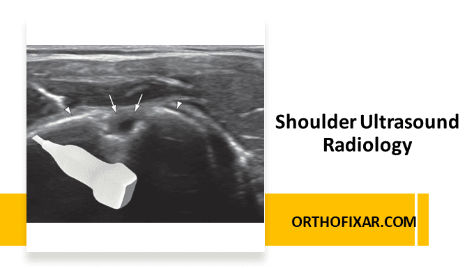
Shoulder ultrasound imaging modality allows for real-time, dynamic evaluation of shoulder structures and provides excellent visualization of soft tissue anatomy. Ultrasound imaging can be used to observe the long head of biceps, the acromiohumeral distance, the subacromial/subdeltoid bursa, the amount of joint laxity, and the rotator cuff anatomy including subscapularis, supraspinatus, infraspinatus and teres minor, for both normal appearances and pathological changes.
The advantages of shoulder ultrasound include its non-invasive nature, cost-effectiveness, lack of ionizing radiation, and ability to perform dynamic assessment during patient movement.
Shoulder Ultrasound Techniques
Anterior View
Long Head of Biceps Ultrasound
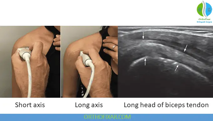
The ultrasound examination for the long head of biceps is done with the patient’s arm is placed in supination with the palm facing upward and the arm resting comfortably on the patient’s own leg. The transducer is initially placed in the transverse plane along the long head of biceps to visualize the tendon in short axis view.
Proper transducer positioning is crucial for optimal visualization. When the transducer is positioned correctly and perpendicular to the tendon, a hyperechoic and well-defined humeral cortex becomes visible underneath the biceps tendon. The tendon should be systematically examined from proximal to distal aspects and will appear as a fibrillary structure presenting as a high-intensity, hyperechoic round, tubular-shaped structure. The biceps tendon maintains a characteristic fibrillar appearance in healthy individuals.
See Also: Long Head of Biceps Tendon
Particular attention should be paid to the tendon’s anatomical placement between the greater and lesser tuberosities within the bicipital groove, which creates a thin hyperechoic rim just deep to the tendon. Recent research has demonstrated that effusion around the long head of biceps shows a moderate to high degree of correlation with range of motion limitations, though it correlates less strongly with functional scores and visual analogue scale measurements for pain in shoulder pathology.
To obtain longitudinal views, the ultrasound transducer should be rotated 90 degrees to visualize the long axis of the tendon. This orientation allows visualization of the tendon from its proximal attachment at the humeral head extending distally near the pectoralis major tendon insertion. Due to the acoustic phenomenon of anisotropy, the transducer may require subtle toggling or rocking movements back and forth along the width of the tendon to achieve optimal visualization in the longitudinal plane.
Acromioclavicular Joint Ultrasound
Assessment of the acromioclavicular joint begins by positioning the transducer superiorly in the transverse plane or short axis orientation over the distal acromioclavicular joint. During examination, either a hypoechoic joint space or a hyperechoic joint disc may be visualized. The cortical outlines of both the acromion and clavicle should be clearly identified.
Through careful visualization of the acromion and clavicle, the acromioclavicular space can be measured, and the hyperechoic interposed fibrocartilage disc can be identified when present. Joint space widening may occur in patients with acromioclavicular joint separation injuries, particularly in grade 3 sprains. When such disruption exists, the distal end of the clavicle will demonstrate elevation with active movement, which can be observed dynamically as the patient moves their arm from a “hand on knee” position to “hand on opposite shoulder” position.

Acromiohumeral Interval Measurement
The acromiohumeral interval distance represents a critical measurement taken between the inferior surface (most lateral edge) of the acromion and the superior surface of the humeral head. This measurement holds significant clinical importance, as decreased acromiohumeral interval distance has been associated with poor surgical outcomes, large rotator cuff tears, and superior humeral migration patterns.
To obtain this measurement, the transducer should be positioned in a coronal oblique plane vertically along the most lateral aspect of the acromion. This positioning allows clear visualization of both the acromion and the greater tuberosity of the humerus, facilitating accurate distance measurement.
Subacromial/Subdeltoid Bursa Evaluation
Using the same transducer position established for acromioclavicular joint assessment, the subacromial/subdeltoid bursa can be effectively evaluated by moving the transducer laterally. The bursa is located directly superior to the supraspinatus muscle and typically appears as a small anechoic rim, which indicates a collapsed subacromial bursa in the resting state.
Dynamic assessment proves particularly valuable when evaluating bursal pathology. When the patient actively moves the shoulder into abduction, pooling of subacromial fluid may become apparent as bursal enlargement, suggesting inflammatory processes or impingement syndromes.
Rotator Cuff Muscle Assessment
Subscapularis Tendon Ultrasound
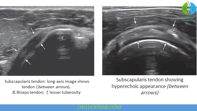
Examination of the subscapularis tendon requires positioning the shoulder in lateral rotation. The initial transducer placement should be in long axis orientation, parallel to the tendon fibers. The tendon should demonstrate hyperechoic characteristics and can be traced laterally to its insertion on the lesser tubercle of the humerus.

Comprehensive examination requires viewing the tendon in its entirety from superior to inferior aspects, with particular emphasis placed on the superior insertion site, as this location represents the most commonly injured area in association with supraspinatus rotator cuff tears. Rotating the transducer 90 degrees allows visualization of the subscapularis tendon along its short axis. Just above the outline of the humeral head, a transverse cross-section of the subscapularis becomes visible. It is normal to observe areas of hypoechoic striations representing muscle tissue or interfaces between the several tendon bundles.
See Also: Subscapularis Muscle Anatomy
Supraspinatus Ultrasound
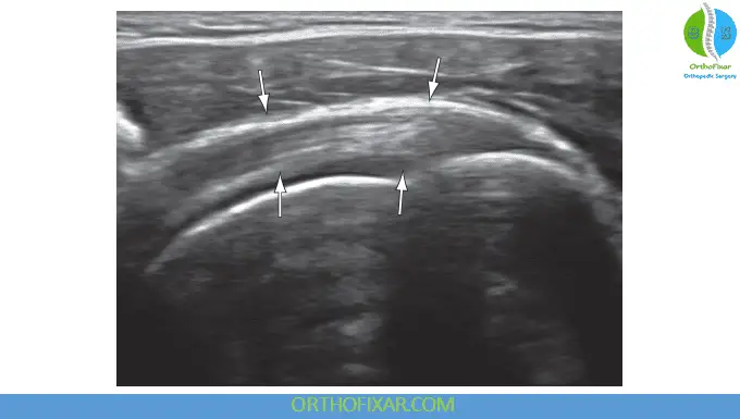
The supraspinatus represents the most commonly torn rotator cuff tendon, making its thorough evaluation critical during any shoulder ultrasound examination. Patient positioning for supraspinatus assessment involves placing the hand of the affected shoulder on the ipsilateral hip, a position known as the “modified Crass” position. This positioning extends the humerus, moving both the greater tuberosity and the supraspinatus tendon from underneath the acromion to expose them anteriorly for optimal visualization.
For longitudinal visualization of the supraspinatus, the transducer should be placed parallel to the supraspinatus fibers in alignment with the ipsilateral humerus. The transducer can be systematically moved both anteromedially and posterolaterally to examine the tendon comprehensively from anterior to posterior aspects. Normal tendon will demonstrate hyperechoic and fibrillary characteristics throughout its length.
Short axis visualization requires rotating the transducer 90 degrees, which positions it perpendicular to the long axis of the ipsilateral humerus. In this view, a hyperechoic rim of the humerus lies beneath the hypoechoic articular cartilage, with the supraspinatus positioned just superior to the hypoechoic articular cartilage. The anterior border of the supraspinatus can be identified just lateral to the long head of biceps tendon.
Posterior View Assessment
Infraspinatus Ultrasound
Posterior assessment focuses primarily on infraspinatus visualization. Optimal patient positioning involves seating the patient with the shoulder in medial rotation and the ipsilateral hand and forearm resting on the patient’s medial thigh. The transducer should be placed longitudinally parallel with the tendon fibers just below the scapular spine.
The infraspinatus tendon fibers appear hyperechoic while positioned above the humeral head and beneath the deltoid muscle fibers. The examination should follow the fibers laterally to their attachment site on the greater tuberosity. Transverse plane visualization of the infraspinatus can be achieved by rotating the transducer 90 degrees so that it runs parallel to the posterior humerus.
Advanced Applications
Humeral Torsion Assessment
Shoulder ultrasound can be utilized to determine humeral torsion, which represents the relative difference in osseous rotation between the proximal and distal articular surfaces of the humerus. This measurement significantly influences shoulder range of motion capabilities. Ultrasonography determines humeral torsion by aligning the apices of the greater and lesser tuberosities and measuring the corresponding forearm angle, providing valuable information for treatment planning and rehabilitation protocols.
Shoulder Ultrasound Clinical Applications
Shoulder ultrasound excels in identifying various pathological conditions including rotator cuff tears, tendinopathy, bursitis, biceps tendon pathology, and joint effusions. The dynamic nature of ultrasound examination allows for assessment of impingement syndromes and evaluation of tendon movement patterns during active motion.
Common pathological findings include tendon thickening, hypoechoic areas within tendons suggesting degeneration, complete or partial thickness tears appearing as anechoic or hypoechoic defects, and bursal thickening or fluid accumulation indicating inflammatory processes.
References & More
- Orthopedic Physical Assessment by David J. Magee, 7th Edition.
- Singh JP. Shoulder ultrasound: What you need to know. Indian J Radiol Imaging. 2012 Oct;22(4):284-92. doi: 10.4103/0971-3026.111481. PMID: 23833420; PMCID: PMC3698891. Pubmed
- Amoo-Achampong K, Nwachukwu BU, McCormick F. An orthopedist’s guide to shoulder ultrasound: a systematic review of examination protocols. Phys Sportsmed. 2016;44(4):407–416. Pubmed
- Lee HJ, Bae SH, Lee KY, et al. Evaluation of the effusion within biceps long head tendon sheath using ultrasonography. Clin Orthop Surg. 2015;7(3): 351–358. Pubmed
- Gaitini D. Shoulder ultrasonography: performance and common findings. J Clin Imaging Sci. 2012;2(1):38. Pubmed
- Seitz AL, Michener LA. Ultrasonographic measures of subacromial space in patients with rotator cuff disease: a systematic review. J Clin Ultrasound. 2011;39(3):146–154. Pubmed
- Bailey LB, Beattie PF, Shanley E, et al. Current rehabilitation applications for shoulder ultrasound imaging. J Ortho Sports Phys Ther. 2015;45(5): 394–405. Pubmed
- Azzoni R, Cabitza P, Parrini M. Sonographic evaluation of subacromial space. Ultrasonics. 2004;42(1-9): 683–687. Pubmed
- Deseules F, Minville L, Riederer B, et al. Acromiohumeral distance variation measured by ultrasonography and its association with the outcome of rehabilitation for shoulder impingement syndrome. Clin J Sport Med. 2004;14(4):197–205. Pubmed
- Jacobson JA: Fundamentals of musculoskeletal ultrasound, ed 3, Philadelphia, 2018, Elsevier
