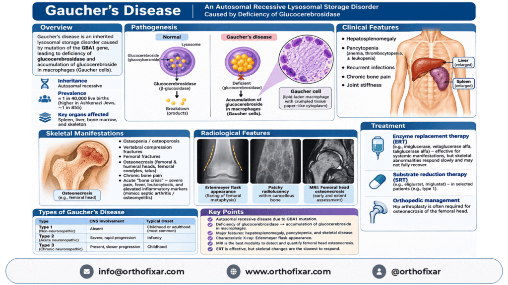
Gaucher disease is a rare, inherited lysosomal storage disorder caused by a deficiency of the enzyme glucocerebrosidase (acid beta-glucosidase). This enzyme normally breaks down glucosylceramide, a lipid derived from the turnover of cell membranes. When the enzyme is deficient, glucosylceramide accumulates within macrophages, producing characteristic Gaucher cells.
Gaucher disease primarily affects the spleen, liver, bone marrow, and skeletal system. Common manifestations include hepatosplenomegaly, anemia, thrombocytopenia, chronic bone pain, osteopenia, pathological fractures, and osteonecrosis.
The condition is inherited in an autosomal recessive pattern and is particularly prevalent among individuals of Ashkenazi Jewish ancestry.
Although Gaucher disease is lifelong, disease-specific therapies can significantly improve many of its systemic manifestations. Early recognition is especially important because some skeletal complications can become irreversible.
What Is Gaucher Disease?
Gaucher disease is a genetic metabolic disorder belonging to the group of lysosomal storage diseases.
Lysosomes are intracellular organelles responsible for breaking down and recycling biological materials. In Gaucher disease, defective lysosomal glucocerebrosidase activity prevents the normal degradation of glucosylceramide, also known as glucocerebroside.
This substance accumulates predominantly within macrophages of the reticuloendothelial system.
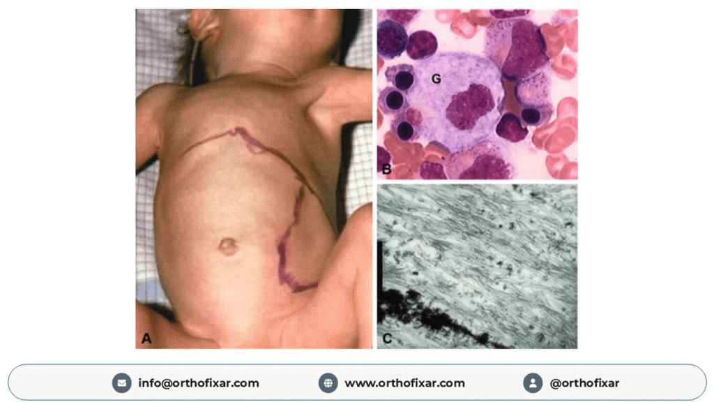
The enlarged, lipid-laden macrophages are called Gaucher cells. Their accumulation contributes to enlargement and dysfunction of affected organs.
The disease has a broad clinical spectrum, ranging from asymptomatic individuals to severe neurological disease beginning in infancy.
Causes and Genetics of Gaucher Disease
GBA1 Gene Mutation
Gaucher disease results from pathogenic variants affecting both copies of the GBA1 gene, located on chromosome 1.
The GBA1 gene encodes lysosomal glucocerebrosidase, an enzyme responsible for hydrolyzing glucosylceramide into glucose and ceramide.
Under normal circumstances, glucosylceramide is generated during the breakdown of glycosphingolipid-rich cell membranes, including those of aging blood cells.
When glucocerebrosidase activity is severely reduced, glucosylceramide cannot be efficiently degraded.
The resulting accumulation contributes to cellular dysfunction, inflammation, and progressive tissue damage.
Autosomal Recessive Inheritance
Gaucher disease follows an autosomal recessive inheritance pattern.
An affected individual usually inherits one pathogenic GBA1 variant from each parent.
When both parents are carriers, the probabilities for each pregnancy are:
- 25%: The child inherits both pathogenic variants and is affected.
- 50%: The child inherits one pathogenic variant and is a carrier.
- 25%: The child inherits neither familial pathogenic variant.
These probabilities apply independently to every pregnancy.
Individuals carrying only one pathogenic GBA1 variant generally do not develop Gaucher disease. However, certain GBA1 variants are associated with an increased risk of Parkinson disease, an important consideration during genetic counseling.
Epidemiology and Prevalence
Gaucher disease is uncommon in the general population.
An approximate birth incidence of 1 in 40,000 live births is often cited, although epidemiological estimates vary considerably by country, population, and study methodology.
Importantly, birth incidence and population prevalence are different measures and should not be used interchangeably.
Gaucher disease is substantially more common in people of Ashkenazi Jewish ancestry.
According to NCBI GeneReviews, type 1 Gaucher disease has an estimated prevalence of approximately 1 in 855 in the Ashkenazi Jewish population, with an estimated carrier frequency of 1 in 18.
Pathophysiology of Gaucher Disease
The principal pathological abnormality is the accumulation of glucosylceramide within macrophage lysosomes.
Formation of Gaucher Cells
Defective glucocerebrosidase activity leads to progressive lipid accumulation within macrophages.
These cells become enlarged and develop characteristic cytoplasm that resembles crumpled tissue paper on light microscopy.
Gaucher cells are particularly prominent in:
- Spleen
- Liver
- Bone marrow
- Other macrophage-rich tissues
Their accumulation, together with changes in inflammatory signaling and tissue remodeling, contributes to organ enlargement, cytopenias, and skeletal abnormalities.
Mechanism of Bone Disease
Skeletal involvement is one of the most clinically important aspects of Gaucher disease.
Marrow infiltration by Gaucher cells and disturbances in bone remodeling contribute to reduced bone mineral density and structural abnormalities.
Patients may subsequently develop:
- Osteopenia and osteoporosis
- Bone marrow infiltration
- Bone infarction
- Osteonecrosis
- Pathological fractures
- Chronic bone pain
- Joint deformity and secondary osteoarthritis
Importantly, skeletal disease does not always correlate with the severity of liver enlargement, splenomegaly, or hematological abnormalities.
Types of Gaucher Disease
Gaucher disease is traditionally classified into three major types based on the presence and progression of neurological involvement.
Type 1 Gaucher Disease (Non-Neuronopathic)
Type 1 is the most common form in many populations.
It primarily affects the spleen, liver, blood cells, and bones, without the primary central nervous system manifestations used to define neuronopathic Gaucher disease.
Clinical manifestations include:
- Hepatosplenomegaly
- Anemia and thrombocytopenia
- Easy bruising or bleeding
- Bone pain and bone crises
- Osteopenia and osteoporosis
- Osteonecrosis
- Pathological fractures
Symptoms may develop in childhood or adulthood, and severity varies widely.
Type 2 Gaucher Disease (Acute Neuronopathic)
Type 2 Gaucher disease is a severe, rapidly progressive form that usually becomes apparent during infancy.
Characteristic features include:
- Progressive neurological deterioration
- Feeding and swallowing difficulties
- Abnormal muscle tone
- Brainstem dysfunction
- Eye movement abnormalities
- Hepatosplenomegaly
Type 2 disease carries a poor prognosis, and currently available enzyme replacement therapy does not reverse its severe neurological manifestations.
Type 3 Gaucher Disease (Chronic Neuronopathic)
Type 3 Gaucher disease involves neurological abnormalities that generally progress more slowly than those in type 2.
Clinical findings may include:
- Abnormal horizontal eye movements
- Seizures
- Ataxia or movement abnormalities
- Hepatosplenomegaly
- Skeletal involvement
- Hematological abnormalities
Type 3 disease has substantial clinical variability.
Comparison of Gaucher Disease Types
| Feature | Type 1 | Type 2 | Type 3 |
|---|---|---|---|
| Primary CNS involvement | Absent | Severe, rapid progression | Present, generally slower progression |
| Typical onset | Childhood or adulthood | Infancy | Usually childhood |
| Skeletal involvement | Common | Generally not prominent | Common |
| Hepatosplenomegaly | Common | Common | Common |
| Disease course | Highly variable | Rapidly progressive | Variable, often chronic |
| Response to enzyme replacement | Systemic benefits | Neurological disease does not respond | Systemic benefits; limited neurological effect |
Clinical Features of Gaucher Disease
Clinical manifestations depend on the disease phenotype, extent of organ involvement, and severity.
Hepatosplenomegaly
Hepatosplenomegaly is a hallmark manifestation of Gaucher disease.
The spleen may become markedly enlarged because of the accumulation of lipid-laden macrophages.
Patients may experience abdominal distension, discomfort, or early satiety.
Splenomegaly may contribute to hypersplenism, resulting in peripheral blood cell sequestration and cytopenias.
The liver may also enlarge and develop variable degrees of involvement.
Hematological Manifestations
Gaucher disease commonly causes anemia and thrombocytopenia.
Anemia may result in fatigue, weakness, and reduced exercise tolerance.
Thrombocytopenia can lead to easy bruising, epistaxis, gum bleeding, and prolonged bleeding.
Leukopenia may also occur, and some individuals develop pancytopenia.
Cytopenias arise through several mechanisms, including hypersplenism and bone marrow involvement.
Infections may occur, but recurrent infections are not a universal or defining manifestation of Gaucher disease.
Other Clinical Manifestations
Depending on the subtype and severity, additional manifestations may include delayed growth, delayed puberty, pulmonary involvement, gallstones, and neurological abnormalities.
Orthopedic Manifestations of Gaucher Disease
Skeletal involvement can be the most disabling component of Gaucher disease.
Patients may develop significant bone abnormalities even when their hematological and visceral manifestations are relatively mild.
Chronic Bone Pain and Joint Stiffness
Chronic bone pain is a frequent complaint, particularly in the lower limbs, pelvis, and spine.
Pain may arise from marrow infiltration, bone infarction, osteonecrosis, or fractures.
Joint stiffness and reduced range of movement may develop, particularly when osteonecrosis progresses to joint collapse and secondary osteoarthritis.
Osteopenia and Osteoporosis
Gaucher disease is associated with decreased bone mineral density.
Generalized osteopenia can increase susceptibility to fragility fractures.
Common complications include:
- Vertebral compression fractures
- Femoral fractures
- Other pathological or fragility fractures
Vertebral compression fractures may cause chronic back pain, loss of height, and spinal deformity.
Osteonecrosis (Avascular Necrosis)
Osteonecrosis is a major skeletal complication of Gaucher disease.
It occurs when bone tissue becomes ischemic and undergoes necrosis.
Frequently affected anatomical sites include:
- Femoral head
- Humeral head
- Femoral condyles
- Talus
Osteonecrosis of the femoral head may produce progressive hip pain, restricted mobility, and eventual articular collapse.
Advanced disease may require total hip arthroplasty.
Bone Infarction
Bone infarction is another important manifestation of Gaucher disease.
Infarction can involve the metaphyseal and diaphyseal regions of long bones.
Patients may experience acute or chronic pain, and imaging can reveal areas of marrow damage.
Bone Crisis in Gaucher Disease
A bone crisis is an acute episode of severe bone pain associated with Gaucher disease.
It may be accompanied by local swelling, tenderness, and systemic inflammatory manifestations.
Clinical Features of Bone Crisis
Typical findings include:
- Sudden onset of severe localized bone pain
- Marked tenderness
- Restricted movement of the affected limb
- Fever or pyrexia
- Occasionally, leukocytosis
- Elevated inflammatory markers
The pain may be severe enough to prevent walking or normal limb movement.
Bone Crisis vs. Osteomyelitis
One of the most important clinical challenges is differentiating a Gaucher bone crisis from an acute bone or joint infection.
Both conditions may present with severe pain, fever, swelling, and inflammatory-marker elevation.
MRI may demonstrate marrow edema and associated abnormalities, but imaging findings can overlap.
A bone crisis must never be assumed to be sterile simply because a patient has Gaucher disease.
Suspected osteomyelitis or septic arthritis requires prompt clinical assessment. Blood cultures, MRI, and joint aspiration may be indicated according to the clinical presentation.
Septic arthritis is particularly time-sensitive because delayed treatment can cause irreversible joint damage.
Radiological Features of Gaucher Disease
Imaging is essential for evaluating skeletal involvement, identifying complications, and monitoring treatment.
Plain Radiographs (X-Rays)
Plain radiographs may demonstrate:
- Patchy radiolucent areas within cancellous bone
- Generalized osteopenia
- Cortical thinning
- Osteosclerosis
- Metaphyseal widening
- Pathological fractures
- Advanced osteonecrosis
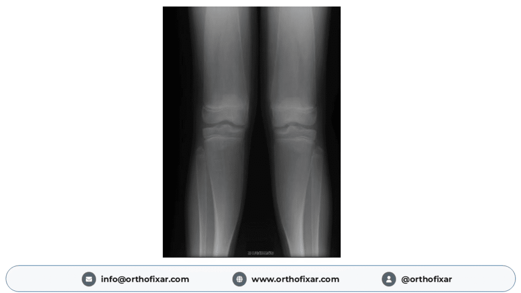
A characteristic skeletal abnormality is the Erlenmeyer flask deformity.
Erlenmeyer Flask Deformity
The Erlenmeyer flask deformity describes abnormal widening and loss of normal metaphyseal contour, most characteristically at the distal femur.
This results from impaired normal bone modeling.
The distal femoral metaphysis becomes broadened, giving the bone a flask-shaped appearance.
Although classically associated with Gaucher disease, the finding is not pathognomonic and may occur in other skeletal or metabolic conditions.
Magnetic Resonance Imaging (MRI)
MRI is particularly valuable for assessing bone marrow involvement and detecting osteonecrosis.
MRI can identify:
- Bone marrow infiltration
- Bone infarction
- Early osteonecrosis
- Marrow edema
- Structural complications
It may detect abnormalities before significant changes become visible on plain radiographs.
MRI is especially useful for evaluating the femoral head when avascular necrosis is suspected.
The extent of osteonecrosis and associated joint involvement can help guide orthopedic management.
Bone Mineral Density Assessment
Dual-energy X-ray absorptiometry (DXA) is used to evaluate bone mineral density.
It is useful in assessing osteopenia and osteoporosis but cannot replace MRI for detecting marrow infiltration or focal osteonecrosis.
Diagnosis of Gaucher Disease
Diagnosis is based on clinical suspicion followed by specific biochemical and/or genetic confirmation.
When Should Gaucher Disease Be Suspected?
Gaucher disease should be considered in patients presenting with combinations of:
- Unexplained hepatosplenomegaly
- Persistent thrombocytopenia or anemia
- Chronic bone pain
- Recurrent episodes of acute bone pain
- Unexplained osteonecrosis
- Pathological fractures
- Characteristic radiological findings
- A relevant family history
Glucocerebrosidase Enzyme Activity Testing
Measurement of glucocerebrosidase activity in peripheral blood leukocytes is a key diagnostic investigation.
Affected individuals typically have markedly reduced enzyme activity, often within approximately 0–15% of normal activity.
Dried blood spot tests can be used in screening, but abnormal screening results require appropriate confirmatory investigations.
Molecular Genetic Testing
Molecular testing identifies pathogenic variants in the GBA1 gene.
Finding pathogenic or likely pathogenic variants in both copies of GBA1 supports or establishes the molecular diagnosis.
Genetic testing is valuable for family counseling, identifying at-risk relatives, and reproductive planning.
Biomarker Testing
Biomarkers can help assess disease burden and treatment response.
Important biomarkers include:
- Glucosylsphingosine (lyso-Gb1)
- Chitotriosidase
- CCL18/PARC
Lyso-Gb1 is particularly useful because of its relatively high diagnostic specificity and value in disease monitoring.
Is Bone Marrow Biopsy Necessary?
Bone marrow biopsy is not routinely required to diagnose Gaucher disease when appropriate enzyme and molecular tests are available.
Although Gaucher cells have characteristic microscopic features, similar-appearing cells may occur in other conditions.
Treatment of Gaucher Disease
Treatment aims to reduce pathological substrate accumulation, improve clinical manifestations, prevent complications, and preserve long-term organ and skeletal function.
Management depends on the Gaucher phenotype, symptom severity, age, and individual clinical circumstances.
Enzyme Replacement Therapy (ERT)
Enzyme replacement therapy is an established treatment for the systemic manifestations of Gaucher disease.
ERT supplies functional glucocerebrosidase to help clear accumulated substrate from affected tissues.
Available enzyme replacement preparations include:
- Imiglucerase
- Velaglucerase alfa
- Taliglucerase alfa
These therapies are given by intravenous infusion according to individualized regimens and applicable approvals.
ERT can improve:
- Hemoglobin concentration
- Platelet counts
- Hepatomegaly
- Splenomegaly
- Bone pain
- Frequency of bone crises
- Some measures of skeletal health
However, skeletal abnormalities often respond more slowly than hematological and visceral manifestations.
Established bone infarction, osteonecrosis, and advanced structural damage may not be reversible.
ERT does not effectively cross the blood-brain barrier and therefore does not adequately treat the primary neurological manifestations of neuronopathic Gaucher disease.
Substrate Reduction Therapy (SRT)
Substrate reduction therapy decreases glucosylceramide synthesis, reducing the amount of material requiring degradation.
Examples include:
- Eliglustat: An oral treatment option for eligible adults with type 1 Gaucher disease. Its use depends on factors including CYP2D6 metabolizer status, contraindications, and drug interactions.
- Miglustat: An oral option for selected patients with type 1 disease when enzyme replacement is unsuitable, subject to local indications.
Treatment selection requires specialist assessment.
Orthopedic Management
Orthopedic treatment is important for patients with significant skeletal complications.
Management may include pain control, rehabilitation, osteoporosis care, fracture management, and surgery when required.
Management of Osteonecrosis
Treatment depends on the affected joint and stage of osteonecrosis.
Early disease may be managed with individualized nonoperative or joint-preserving strategies.
Advanced femoral head osteonecrosis with collapse and disabling pain may require total hip arthroplasty.
Management of Fractures
Fracture treatment follows orthopedic principles while accounting for underlying bone quality, bleeding risk, and disease severity.
Patients may require immobilization, surgical fixation, rehabilitation, or management of associated osteoporosis.
Supportive Treatment
Additional supportive care may include:
- Appropriate calcium and vitamin D intake
- Management of osteoporosis
- Pain control
- Physiotherapy and rehabilitation
- Monitoring of blood counts
- Nutritional and growth assessment in children
- Genetic counseling
- Multidisciplinary specialist follow-up
Monitoring and Long-Term Follow-Up
Gaucher disease requires ongoing clinical monitoring, including patients receiving effective treatment.
Follow-up may involve:
- Complete blood count and platelet count
- Liver and spleen volume assessment
- Lyso-Gb1 or other appropriate biomarkers
- Assessment of bone pain and mobility
- MRI when indicated for skeletal involvement
- DXA for bone mineral density
- Neurological assessment when clinically appropriate
Monitoring intervals should be individualized according to age, disease type, severity, and response to treatment.
Prognosis of Gaucher Disease
The prognosis varies substantially between disease types.
Patients with type 1 Gaucher disease may experience significant improvements in hematological parameters, organ enlargement, symptoms, and quality of life with appropriate treatment.
Nevertheless, irreversible skeletal complications can continue to cause disability.
Type 2 Gaucher disease is associated with rapidly progressive neurological deterioration and a poor prognosis.
Type 3 disease has a variable course, with chronic neurological and systemic involvement.
Early diagnosis and appropriate disease-specific treatment are important for preventing avoidable complications.
Important Examination Points About Gaucher Disease
For medical students and orthopedic trainees, the following findings are particularly important:
- Inheritance: Autosomal recessive.
- Gene involved: GBA1.
- Enzyme deficiency: Lysosomal glucocerebrosidase (acid beta-glucosidase).
- Accumulated substrate: Glucosylceramide (glucocerebroside).
- Characteristic pathological cell: Gaucher cell, with crumpled tissue paper-like cytoplasm.
- Common systemic features: Hepatosplenomegaly, anemia, and thrombocytopenia.
- Major orthopedic complications: Osteopenia, fractures, bone infarction, and osteonecrosis.
- Characteristic X-ray finding: Erlenmeyer flask deformity of the distal femoral metaphysis.
- Important acute presentation: Bone crisis, which may mimic osteomyelitis.
- Important imaging modality: MRI for marrow infiltration and osteonecrosis.
- Confirmatory investigations: Glucocerebrosidase enzyme activity and GBA1 molecular testing.
- Disease-specific treatment: Enzyme replacement therapy or substrate reduction therapy in appropriate patients.
Frequently Asked Questions About Gaucher Disease
What is the main cause of Gaucher disease?
Gaucher disease is caused by pathogenic variants affecting both copies of the GBA1 gene, resulting in deficient lysosomal glucocerebrosidase activity and accumulation of glucosylceramide.
Is Gaucher disease autosomal dominant or autosomal recessive?
Gaucher disease is inherited in an autosomal recessive manner. An affected individual usually inherits a disease-causing GBA1 variant from each parent.
Which enzyme is deficient in Gaucher disease?
The deficient enzyme is glucocerebrosidase, also called acid beta-glucosidase or glucosylceramidase.
What accumulates in Gaucher disease?
Glucosylceramide, also called glucocerebroside, accumulates primarily within macrophage lysosomes.
What is the most characteristic X-ray finding in Gaucher disease?
The Erlenmeyer flask deformity is a classic skeletal finding, characterized by abnormal widening and loss of the normal contour of the distal femoral metaphysis.
Why does Gaucher disease cause osteonecrosis?
Osteonecrosis is associated with complex changes in bone marrow infiltration, inflammation, bone remodeling, and vascular function that can impair bone viability.
Can Gaucher disease cause hip replacement?
Yes. Severe osteonecrosis of the femoral head can lead to articular collapse and secondary arthritis. Total hip arthroplasty may be required when symptoms and structural damage become advanced.
Can Gaucher disease be cured?
Currently available enzyme replacement and substrate reduction therapies can control many systemic manifestations but are not established cures for the underlying inherited condition.
What is the difference between Gaucher disease and Niemann-Pick disease?
Both are inherited lysosomal storage disorders, but they differ in the affected molecular pathways. Gaucher disease is associated with deficient glucocerebrosidase activity and glucosylceramide accumulation, whereas Niemann-Pick diseases involve other defects in sphingolipid metabolism or intracellular cholesterol trafficking.
Conclusion
Gaucher disease is an autosomal recessive lysosomal storage disorder caused by GBA1 mutations and deficient glucocerebrosidase activity.
The accumulation of glucosylceramide in macrophages contributes to hepatosplenomegaly, hematological abnormalities, and significant skeletal complications.
Orthopedic manifestations are particularly important because patients may develop chronic bone pain, osteopenia, pathological fractures, bone infarction, and osteonecrosis.
The Erlenmeyer flask deformity is a classic radiographic feature, while MRI is particularly useful for assessing bone marrow involvement and identifying osteonecrosis.
Although enzyme replacement therapy has substantially improved the management of Gaucher disease, skeletal complications can be slow to respond and may become irreversible.
Early recognition, biochemical confirmation, genetic evaluation, appropriate disease-specific treatment, and multidisciplinary follow-up are essential for optimizing long-term outcomes.
Medical References
The following sources support the genetic, clinical, imaging, and management information discussed in this article.
- Hughes DA, Pastores GM. Gaucher Disease. GeneReviews®. NCBI Bookshelf. Updated December 7, 2023. Read on NCBI
- Hughes D, et al. Gaucher Disease in Bone: From Pathophysiology to Practice. Journal of Bone and Mineral Research. 2019;34(6):996–1013. PubMed PMID: 31233632
- Nalysnyk L, et al. Gaucher disease epidemiology and natural history: a comprehensive review of the literature. Hematology. 2017;22(2):65–73. PubMed PMID: 27762169
- Biegstraaten M, et al. Management goals for type 1 Gaucher disease: An expert consensus document from the European working group on Gaucher disease. Blood Cells, Molecules, and Diseases. 2018;68:203–208. PubMed PMID: 28274788
- Skeletal aspects of Gaucher disease: a review. British Journal of Radiology. 2002. PubMed PMID: 12036828
- Challenges in Gaucher disease: Perspectives from an expert panel. 2025. PubMed PMID: 40112481