When studying medical imaging, understanding the differences between CT and MRI is essential for any student entering radiology, medicine, or allied health fields. CT (computed tomography) and MRI (magnetic resonance imaging) are two of the most widely used cross‑sectional imaging modalities, yet they operate on very different physical principles, each with unique strengths, limitations, and clinical indications. This guide breaks down the core concepts of CT vs MRI, helps you decide which study to order, and gives you practical tips for image interpretation. By the end, you will have a clear, up‑to‑date framework that you can apply in exams and clinical rotations.
Understanding the Basics: CT and MRI Defined
- CT uses X‑rays to produce detailed cross‑sectional images of the body. The X‑ray tube rotates around the patient, and detectors measure the amount of radiation that passes through tissues. Different tissues attenuate X‑rays to varying degrees, which creates contrast in the image.
- MRI uses a strong magnetic field and radiofrequency pulses to align and then disturb hydrogen protons in the body. As the protons relax back to their original state, they emit signals that are processed into images. MRI does not use ionizing radiation.
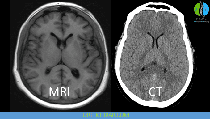
- CT is excellent for evaluating bone, acute bleeding, and calcifications. It is fast – a typical scan takes seconds to a few minutes.
- MRI provides superior soft‑tissue contrast, making it ideal for brain, spinal cord, joints, muscles, and the female pelvis. A routine MRI can take 20 to 45 minutes.
Key Differences Between CT and MRI
The table below summarises the most important parameters when comparing CT vs MRI. Use this as a quick reference during your studies.
| Parameter | CT | MRI |
|---|---|---|
| Radiation | Ionising radiation (X‑rays) | No ionising radiation |
| Scan time | Seconds to a few minutes | 20–45 minutes (or longer) |
| Soft‑tissue contrast | Good for lung, bone, fat, and fluid; poor for subtle soft‑tissue differences | Excellent – can distinguish grey/white matter, tumours, inflammation, and oedema |
| Bone imaging | Best for cortical bone, fractures, and calcifications | Bone appears dark (low signal); better for bone marrow pathology |
| Contrast agents | Iodinated contrast (risk of allergic reaction and nephrotoxicity) | Gadolinium‑based contrast (risk of nephrogenic systemic fibrosis in severe renal impairment) |
| Cost | Lower (typically $300–$1,000) | Higher (typically $600–$2,500) |
| Patient experience | Open, quiet scanner; less claustrophobia | Narrow bore, loud knocking noise; claustrophobia is a common issue |
| Metallic implants | Generally safe (except for some metallic objects that cause streak artefact) | Strict contraindication for many metal implants (aneurysm clips, pacemakers, cochlear implants) |
Clinical Indications: When to Use CT vs MRI
Choosing the right modality depends on the clinical question. Here are the most common scenarios where CT is preferred, and where MRI becomes the better choice.
When CT Is Indicated
- Trauma: CT is the first‑line for acute head injury, facial fractures, cervical spine fractures, and whole‑body trauma (polytrauma).
- Stroke (acute): Non‑contrast head CT rules out intracranial haemorrhage before giving thrombolytics.
- Pulmonary embolism: CT pulmonary angiography (CTPA) is the gold standard.
- Acute abdomen: CT with contrast is used for suspected appendicitis, diverticulitis, bowel obstruction, or kidney stones.
- Lung imaging: CT is best for detecting small pulmonary nodules, interstitial lung disease, and pneumonia complications.
- Bone: CT is ideal for complex fractures, especially of the pelvis, acetabulum, and calcaneus.
When MRI Is Indicated
- Brain and spinal cord: MRI is superior for multiple sclerosis, brain tumours, spinal cord compression, and demyelinating diseases.
- Joints and soft tissues: Meniscal tears, rotator cuff injuries, ligament tears, and cartilage defects are best seen on MRI.
- Female pelvis: MRI is excellent for evaluating uterine fibroids, endometriosis, and ovarian lesions.
- Prostate cancer: Multiparametric MRI is now standard for detection and staging.
- Abdominal and pelvic masses when contrast is contraindicated: MRI can often provide better characterisation without iodine.
- Paediatric imaging: Because MRI has no radiation, it is preferred for imaging children, especially for brain and spinal problems.
“A 45-year-old man arrives in the emergency department with sudden severe headache and loss of consciousness. The clinical suspicion is subarachnoid haemorrhage. The most appropriate first imaging study is a non-contrast CT of the head – it is fast, widely available, and extremely sensitive for acute blood. If the CT is negative but clinical suspicion remains high, a lumbar puncture or MRI may be performed later.”
Image Interpretation: What Students Need to Look For
Learning to read CT and MRI images takes years of practice, but as a student you should focus on basic principles and common patterns.
CT Image Interpretation
- Understand windowing: CT images are viewed in different windows – brain window (level ~40, width ~80), bone window (level ~300, width ~1500), lung window (level ~-600, width ~1500), and abdominal soft‑tissue window (level ~50, width ~350). Each window highlights different tissues.
- Look for symmetry: Compare left and right sides of the brain, chest, and abdomen. Asymmetry often indicates pathology.
- Know the density scale: Air is black (‑1000 HU), fat is dark gray (-50 to -100 HU), water is gray (0 HU), soft tissue is light gray (40–80 HU), bone is white (300+ HU), and calcium is very white (100+ HU).
- Acute blood on CT is moderately hyperdense (about 60–80 HU) and appears white compared to brain tissue.
- Contrast enhancement: Tissues that enhance after intravenous contrast indicate increased blood flow (tumours, infection, inflammation). Lack of enhancement can suggest necrosis or thrombosis.
MRI Image Interpretation
- Understand basic sequences: T1‑weighted images show anatomy well – fluid is dark, fat is bright. T2‑weighted images show pathology – fluid is bright, fat is still relatively bright but less so. FLAIR (Fluid Attenuated Inversion Recovery) suppresses fluid signals so that periventricular lesions become more visible.
- Signal intensity: In T1, isointense to grey matter is typical; in T2, hyperintense lesions often indicate oedema, demyelination, or tumour. Hypointensity on T2 may be due to blood breakdown products (e.g., haemosiderin in old haemorrhage) or calcification.
- Look for mass effect: A large lesion can shift midline structures, compress ventricles, or efface sulci. This is a critical red flag.
- Contrast enhancement in MRI using gadolinium: Enhancement indicates breakdown of the blood‑brain barrier (e.g., in active multiple sclerosis plaques, abscesses, or high‑grade tumours).
- Artifact recognition: Motion artifact appears as blurring, ghosting, or banding. Metal artifact (from braces, joint implants) causes signal loss and distortion. Susceptibility artifact (from haemoglobin breakdown products) causes blooming on GRE/SWI sequences.
“When you see a bright white spot on a T2‑weighted brain MRI in a young woman with episodes of optic neuritis, think about multiple sclerosis. Multiple sclerosis plaques are typically ovoid, periventricular, and perpendicular to the ventricular surface. Follow the ‘Dawson’s fingers’ sign – it is a classic pattern in MRI interpretation.”
Advantages and Limitations
CT Advantages
- Very fast – ideal for unstable patients or uncooperative children.
- Widely available in emergency settings.
- Excellent for bone, lung, and acute haemorrhage.
- Lower cost compared to MRI.
CT Limitations
- Ionising radiation – cumulative risk, especially in young patients and those requiring repeated scans.
- Iodinated contrast can cause allergic reactions and nephrotoxicity.
- Poor soft‑tissue contrast (cannot distinguish tumour from oedema as well as MRI).
- Beam‑hardening artifact from metal or bone can obscure subtle findings.
MRI Advantages
- Superior soft‑tissue contrast – can differentiate subtle differences in tissue composition.
- No ionising radiation – safer for paediatric and pregnant patients (second and third trimesters).
- Multiplanar imaging – direct coronal, sagittal, and axial views without patient repositioning.
- Functional capabilities (diffusion, perfusion, spectroscopy, fMRI).
MRI Limitations
- Long scan times – motion artifact is a major problem.
- Claustrophobia – up to 5% of patients cannot complete the scan.
- Gadolinium can cause nephrogenic systemic fibrosis in patients with severe kidney disease.
- Ferromagnetic implants are contraindicated – strict screening required.
- High cost and limited availability in some regions.
Practical Examples for Students
Here are two common clinical vignettes to apply your knowledge of CT vs MRI.
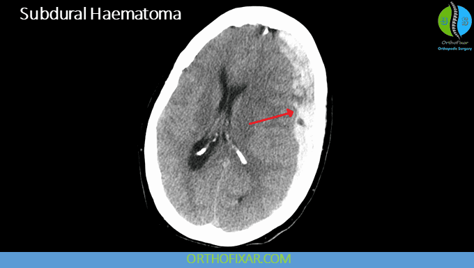
Example 1 – Acute Head Trauma: A 70-year-old man on warfarin fell and hit his head. He is now drowsy with a Glasgow Coma Scale of 13. The emergency doctor orders a non‑contrast head CT. On the CT, you see a hyperdense crescent‑shaped collection along the left convexity, crossing suture lines – this is a subdural haematoma. The CT is fast (less than 5 minutes) and clearly shows the blood, which is hyperdense because of the acute clot. No MRI needed here – the CT gives the surgeon all necessary information for emergency evacuation.
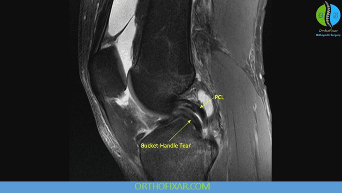
Example 2 – Chronic Knee Pain: A 30-year-old athlete has persistent knee pain and locking after a twisting injury. No fracture on X‑ray. The orthopaedic surgeon orders an MRI of the knee. On the sagittal proton‑density sequence, you see a linear high signal within the medial meniscus that extends to the articular surface – this is a meniscal tear. MRI is the perfect choice because it shows cartilage, ligaments, tendons, and bone marrow oedema with exquisite detail. CT would not adequately visualise the meniscus.
Safety Considerations
- CT: Always check pregnancy status in women of childbearing age. Use the ALARA principle (As Low As Reasonably Achievable) – minimise radiation dose by using paediatric protocols when appropriate.
- MRI: Screen every patient for metal – pacemakers, implanted cardiac devices, aneurysm clips, cochlear implants, shrapnel, and metallic foreign bodies in the eyes. Ferromagnetic objects can become projectiles or cause thermal injury. Also ask about claustrophobia – some patients require sedation or an open MRI.
- Contrast: Both iodinated and gadolinium‑based contrast carry risks. Pre‑medicate for allergic history, check renal function, and be aware of the risk of contrast‑induced nephropathy (CT) and NSF (MRI).
Conclusion
Mastering CT vs MRI is a foundational skill for any medical or radiology student. CT remains the workhorse for trauma, acute emergencies, and lung/bone pathology, while MRI excels in soft‑tissue characterisation, neurological disorders, and joint imaging. By understanding the principles, indications, and basic interpretation tips outlined here, you will be better prepared to select the right study and recognise key findings. Remember that no single modality is always best – the clinical question, patient factors, and available resources should guide your choice. Keep practising with real cases, and soon the differences will become second nature.
Frequently Asked Questions (FAQ)
1. Which imaging modality is better for the brain – CT or MRI?
For most brain pathologies, MRI is superior because of its excellent soft‑tissue contrast. CT is used primarily in the acute setting to rule out haemorrhage, fracture, or mass effect quickly. For chronic conditions like multiple sclerosis, tumours, or dementia, MRI is the preferred test.
2. Can MRI replace CT for all indications?
No. CT is much faster and better for imaging bone, lung parenchyma, and acute haemorrhage. MRI cannot replace CT in trauma or pulmonary embolism. In some situations, such as evaluating kidney stones, CT is also more sensitive.
3. Is CT or MRI more dangerous to the patient?
CT exposes the patient to ionising radiation, which carries a small long‑term cancer risk. MRI has no radiation but has risks related to magnetic fields, contrast agents, and claustrophobia. The overall risk profile depends on the patient’s age, medical condition, and the number of scans.
4. Why does MRI take so much longer than CT?
MRI requires multiple sequences that use different radiofrequency pulses and gradient patterns to obtain various tissue contrasts. Each sequence can take 2–8 minutes, and a complete study often includes 4–8 sequences. CT acquires all slices in a single rotation, usually under 30 seconds.
5. What is the difference between T1 and T2 in MRI?
In T1‑weighted images, fat is bright and water is dark, so anatomy is well visualised. In T2‑weighted images, water is bright, making oedema, inflammation, and most pathology appear bright. Understanding these basic signal differences is the first step in MRI interpretation.
6. When should contrast be used in CT and MRI?
Contrast is used to highlight areas of abnormal blood‑brain barrier breakdown, increased vascularity, or infection. In CT, iodinated contrast helps characterise abscesses, tumours, and vascular lesions. In MRI, gadolinium enhances active inflammation, tumours, and demyelination. Contrast is not needed for simple fractures or routine lung imaging.
7. Can a patient with a pacemaker have an MRI?
Most modern pacemakers are MRI‑conditional under strict protocols, but many older models remain a contraindication. The patient must be screened and the pacemaker interrogated before scanning. In general, MRI is avoided in patients with non‑MRI‑conditional cardiac devices.
8. How do you distinguish between a subdural and epidural haematoma on CT?
On CT, a subdural haematoma appears as a crescent‑shaped hyperdense collection that crosses suture lines but does not cross the falx. An epidural haematoma appears as a biconvex (lentiform) hyperdense collection that does not cross suture lines but can cross the midline at the falx. This difference is critical in emergency assessment.
9. Do CT and MRI use the same contrast agents?
No. CT uses iodinated contrast, which blocks X‑rays and appears white on images. MRI uses gadolinium‑based contrast, which shortens the T1 relaxation time and appears bright on T1‑weighted images. They are chemically different and carry distinct risks.
10. What is the best way for a student to start learning image interpretation?
Start with anatomy – label normal structures on CT and MRI using atlases or online resources. Then learn to recognise common patterns (e.g., fluid levels, mass effect, symmetry). Finally, study cases systematically: identify the modality, the anatomical region, the findings, and the most likely diagnosis. Practice with real anonymised images and ask a senior colleague for feedback.