The Sacroiliac Rocking Test, also known as the Sacrotuberous Ligament Stress Test, is used to assess the integrity and pain response of the sacroiliac (SI) joint and the sacrotuberous ligament. It helps identify dysfunction or irritation within these structures.
The sacroiliac joint is a large, weight-bearing articulation between the sacrum and ilium. The sacrotuberous ligament is a strong, fan-shaped ligament that runs from the ischial tuberosity to the posterior aspect of the ilium and sacrum. This ligament provides stability to the sacroiliac joint and posterior pelvis.
See Also: Pelvic Anatomy: Bone & Ligaments
How to Perform the Sacroiliac Rocking Test?
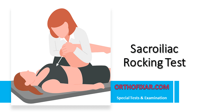
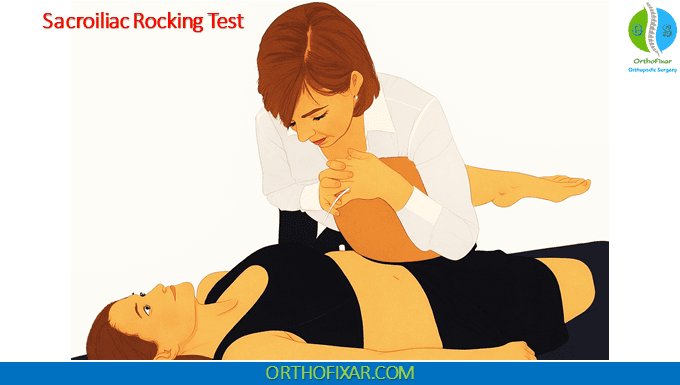
- The patient lies in a supine position on the examination table.
- The examiner flexes the patient’s hip and knee fully.
- The hip is then adducted toward the midline.
- To perform the maneuver correctly, the knee is moved toward the patient’s opposite shoulder, creating a “rocking” motion at the sacroiliac joint.
- Both the hip and knee should have full range of motion and be free from pathology before performing the test.
- Some authors recommend adding medial rotation of the hip during flexion and adduction to further increase stress on the SI joint.
- During the test, the examiner may palpate the sacrotuberous ligament (located between the sacrum and the ischial tuberosity) to check for tenderness or abnormal tension.
Critical Requirement: To perform this test properly, both the hip and knee must demonstrate no pathology and have full range of motion (ROM). The presence of hip or knee pathology will invalidate the test results and may cause unnecessary discomfort to the patient.
See Also: Sacroiliac Joint Dislocation
What does a Positive Sacroiliac Rocking Test Mean?
Pain reproduced in the sacroiliac joint region indicates a positive test and suggests sacroiliac joint dysfunction or ligamentous irritation. The test can also produce tenderness along the sacrotuberous ligament if it is inflamed or stressed.
The Sacroiliac Rocking Test places significant stress on both the hip and sacroiliac joints. It should be performed slowly and carefully, especially in patients with known lower back or hip pathology. If a longitudinal force is applied through the femur in a slow, steady manner (15–20 seconds) in an oblique and lateral direction, additional stress is placed on the sacrotuberous ligament for further assessment.
References & More
- Lee D. The Pelvic Girdle. 2nd ed. Edinburgh: Churchill Livingstone; 1999.
- Porterfield JA, DeRosa C. Mechanical Low Back Pain: Perspectives in Functional Anatomy. Philadelphia: WB Saunders; 1991
- Wong M, Sinkler MA, Kiel J. Anatomy, Abdomen and Pelvis, Sacroiliac Joint. [Updated 2023 Aug 8]. In: StatPearls [Internet]. Treasure Island (FL): StatPearls Publishing; 2025 Jan-. Available from: PubMed
- Orthopedic Physical Assessment by David J. Magee, 7th Edition.
