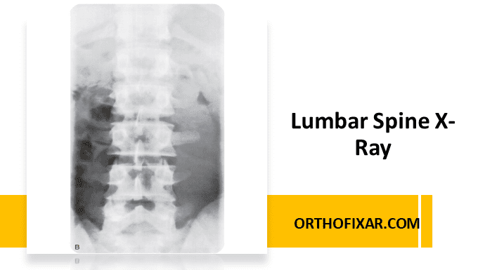
Lumbar spine X-ray (also called lumbar X-ray or lumbosacral spine X-ray) is a common imaging study used to evaluate the bones and alignment of the lower back. It helps detect fractures, degenerative changes, and structural abnormalities of the lumbar vertebrae.
Routine plain lumbosacral X-rays are most appropriate when risk factors for a vertebral fracture are present, or if the patient’s symptoms have not improved after about one month of conservative management. In adults under 50 years of age with no signs or symptoms of systemic disease, imaging is generally not required. For patients over 50 years, lumbar x-ray along with laboratory tests can help rule out most systemic conditions.
See Also: Lumbar Spine Anatomy
Risk Factors for Vertebral Fractures
- Age 50 years or older
- Significant trauma (such as a fall or external injury)
- History of osteoporosis
- Corticosteroid use
- Substance abuse, which increases the likelihood of trauma
See Also: Thoracolumbar Spine Burst Fractures
Standard Lumbar X-Ray Views
Routine Lumbar Spine X-Ray typically includes anteroposterior (AP) and lateral views. In some cases, two lateral views may be taken—one showing the entire lumbar spine and another focusing on the lower two segments. Oblique views are added if spondylolysis or spondylolisthesis is suspected.
Anteroposterior (AP) View
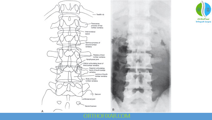
In the AP lumbar vertebrae X-ray, the examiner should assess:
- Shape of the vertebrae — look for any wedging or compression suggesting a fracture.
- Intervertebral disc spaces — assess for reduced height as seen in spondylosis.
- Vertebral deformities — such as hemivertebrae or other congenital anomalies.
- Bamboo spine — characteristic of ankylosing spondylitis.
- Transitional vertebrae:
- Lumbarization of S1 (seen in 2–8% of the population), where S1 behaves like a lumbar vertebra, making S1–S2 the first mobile segment.
- Sacralization of L5 (3–6% incidence), where L5 fuses with the sacrum, making L4–L5 the first mobile level.
- Spina bifida occulta, which may be seen in 6–10% of individuals.
Lateral View
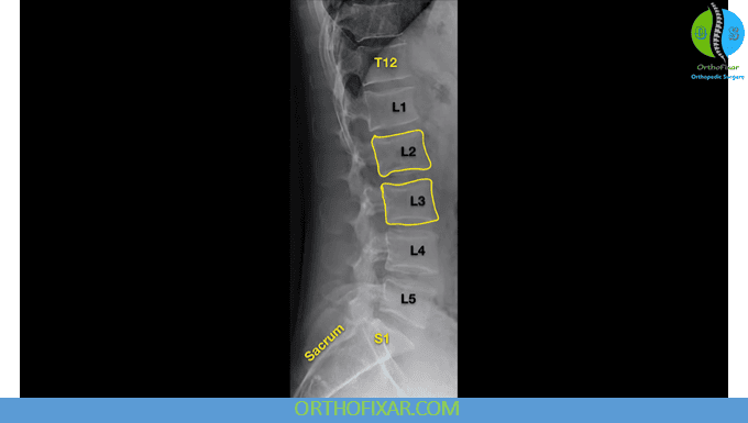
The lateral lumbar X-ray is essential for evaluating alignment, stability, and intervertebral spacing. The examiner should note:
- Spondylosis or spondylolisthesis (found in 2–4% of the population). The degree of vertebral slipping can be graded.
- Lumbar lordosis — normal curvature should be maintained.
- Intervertebral foramina — check for narrowing or distortion.
- Disc spacing — reduced space indicates disc degeneration.
- Alignment of vertebral bodies — loss of the smooth curve suggests spinal instability.
- Osteophyte formation or traction spurs — a traction spur develops about 1 mm from the disc margin and indicates instability, while an osteophyte forms directly at the vertebral margin.
Oblique View
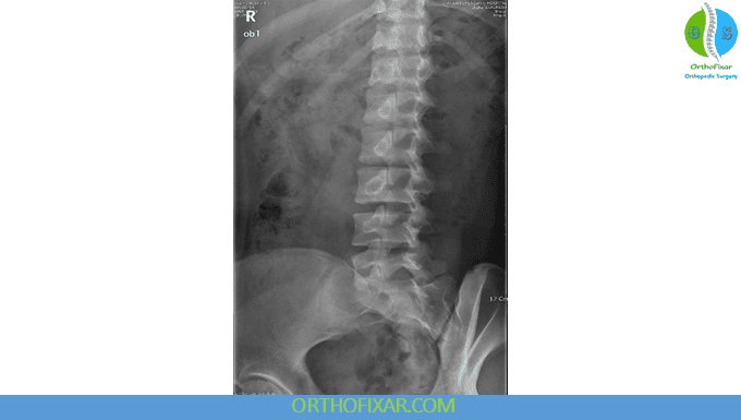
The oblique lumbar X-ray helps detect pars interarticularis defects.
- A “Scottie dog with a collar” appearance indicates spondylolysis.
- A “decapitated Scottie dog” appearance indicates spondylolisthesis.
Motion Views
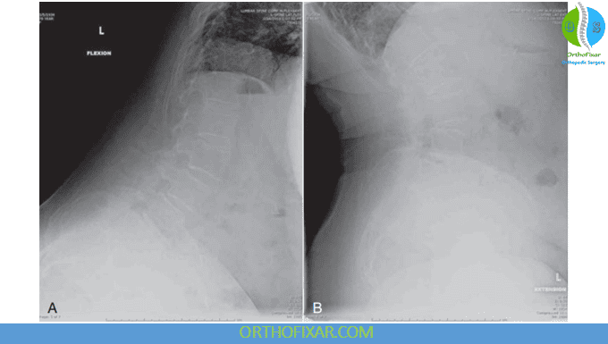
Dynamic lumbar spine X-rays (flexion and extension lateral views) can reveal abnormal spinal motion or instability. These motion views are especially helpful in diagnosing spondylolisthesis or subtle segmental instability. In some cases, anteroposterior side-bending views are also obtained.
Summary
A lumbar spine X-ray remains a valuable diagnostic tool when used appropriately — especially for patients with lower back pain and risk factors for fracture or instability. Proper evaluation of the anteroposterior, lateral, and oblique views provides vital information about vertebral shape, alignment, disc height, and structural abnormalities.
References & More
- Jarvik JG, Deyo RA. Diagnostic evaluation of low back pain with emphasis on imaging. Ann Intern Med. 2002;137:586–597. PubMed
- Deyo RA, Bigos SJ, Maravilla KR. Diagnostic imaging procedures for the lumbar spine. Ann Intern Med. 1989;111:865–868. PubMed
- Li Y, Hresko MY. Radiographic analysis of spondylolisthesis and sagittal spinopelvic deformity. J Am Acad Orthop Surg. 2012;20(4):194–205. PubMed
- Pate D, Goobar J, Resnick D, et al. Traction osteophytes of the lumber spine: radiographic: pathologic correlation. Radiology. 1988;166:843–846. PubMed
- Bigg-Wither G, Kelly P. Diagnostic imaging in musculoskeletal physiotherapy. In: Refshauge K, Gass E, eds. Musculoskeletal Physiotherapy: Clinical Science and Practice. Oxford: Butterworth Heinemann; 1995.
- Wood KB, Popp CA, Transfeldt EE, et al. Radiographic evaluation of instability in spondylolisthesis. Spine. 1994;19:1697–1703. PubMed
- Orthopedic Physical Assessment by David J. Magee, 7th Edition.
