Transient osteoporosis of the hip is a rare but treatable condition in which the bone in your hip joint temporarily loses density and fills with fluid, causing pain that can make walking and standing difficult. The good news is that it is usually self-limiting: with the right diagnosis, protected weight bearing, and a rehabilitation plan, most people recover fully. This article explains what causes it, how it is diagnosed, what treatment actually helps, and what recovery realistically looks like.
What Is Transient Osteoporosis of the Hip?
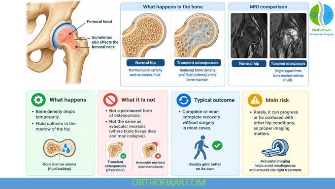
Transient osteoporosis of the hip is a temporary condition affecting the femoral head and sometimes the femoral neck. The bone becomes less dense than normal, and the bone marrow inside the hip fills with edema, which is fluid buildup.
The word “transient” is the key part. Unlike avascular necrosis, where bone tissue dies and may collapse, this condition usually reverses on its own.
It is also known as bone marrow edema syndrome of the hip. Some doctors consider the two terms slightly different, but in everyday practice they describe the same clinical picture.
- What happens: Bone density drops temporarily and fluid collects in the marrow of the hip.
- What it is not: It is not a permanent form of osteoporosis and not the same as avascular necrosis.
- Typical outcome: Complete or near-complete recovery without surgery in most cases.
- Main risk: Rarely, it can progress or be confused with other hip conditions, so proper imaging matters.
Who Develops It and Why
Transient osteoporosis of the hip most often appears in two very different groups of people: middle-aged men and women in the later stages of pregnancy.
In men, it typically shows up between the ages of 40 and 60, often without an obvious trigger. In pregnancy, it usually appears in the third trimester and is linked to the physical and hormonal changes of late pregnancy.
Researchers have proposed several explanations, including temporary disruption of blood flow to the hip, hormonal shifts, mechanical stress, and low vitamin D levels. No single cause has been confirmed.
- Middle-aged men without a clear injury or risk factor.
- Women in the third trimester of pregnancy, sometimes in both hips.
- People with low vitamin D or calcium intake.
- Occasionally people after a minor hip trauma, prolonged immobilization, or with a history of similar episodes.
Is It the Same in Pregnancy?
Pregnancy-related cases tend to be milder and often settle within weeks to a few months after delivery. Treatment is usually conservative, and imaging choices are made with the baby’s safety in mind.
Symptoms to Watch For
The pain of transient osteoporosis of the hip usually comes on gradually. There is often no single moment of injury that you can point to.
Many people describe a deep ache in the groin, front of the thigh, or buttock. The pain gets worse when you stand on the affected leg or walk, and it often eases when you rest.
- Gradual groin or hip pain that builds over days to weeks.
- Pain that worsens with standing, walking, or turning in bed.
- Night pain that can disturb sleep.
- A limp or a feeling that the leg will not take your full weight.
- Reduced hip movement, especially rotating the leg inward.
- Pain that may radiate to the thigh or knee, which sometimes confuses the diagnosis.
A key feature is that the hip is usually not swollen, red, or hot. Fever and severe night sweats point toward infection instead and need urgent assessment.
Transient osteoporosis of the hip feels serious, and it is painful. But the name matters: in most cases, the bone changes are temporary and the joint recovers.
How Doctors Confirm the Diagnosis
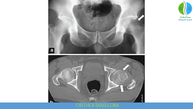
Plain X-rays are often normal in the first few weeks. That is one reason the condition is frequently missed early on.
MRI is the imaging test of choice. It shows the bone marrow edema clearly and helps rule out avascular necrosis, a stress fracture, an infection, or a tumor.
Blood tests help exclude other causes of hip pain, including inflammatory conditions and infection. In pregnancy, your doctor will choose imaging carefully and may avoid contrast agents.
- X-ray: May show reduced bone density in the femoral head later, but can be normal early.
- MRI: The most sensitive test for bone marrow edema and the key to diagnosis.
- Blood tests: Used to rule out infection, inflammation, and metabolic bone disease.
- Follow-up imaging: Sometimes repeated to confirm the changes are resolving.
Conditions That Look Similar
Avascular necrosis is the most important condition to separate from transient osteoporosis of the hip. It has a similar appearance on early scans but a very different long-term course.
Phases of the Condition and What to Expect
The condition generally moves through predictable phases. Knowing them helps you plan work, childcare, and daily routines.
| Phase | Typical Timing | What Is Happening | Practical Focus |
|---|---|---|---|
| Onset | First few weeks | Pain builds, X-ray may be normal | Get an MRI, start offloading the hip |
| Peak | Weeks 4 to 8 | Pain is most intense, walking is hardest | Crutches, rest, pain control, gentle range of motion |
| Regression | Months 3 to 6 | Bone marrow edema starts to shrink on MRI | Progressively increase protected activity and physiotherapy |
| Recovery | Months 6 to 12 | Bone density returns, symptoms fade | Return to normal walking, sport, and work |
Timelines vary widely. Some people improve within three months, while others take a year or more to feel completely normal.
Treatment Options That Actually Help
There is no single drug that cures transient osteoporosis of the hip. Treatment focuses on protecting the hip while the bone heals itself.
Protected weight bearing is the foundation. That usually means using crutches or a walker so the affected hip carries as little load as possible during the painful phase.
- Offloading: Crutches, a cane, or a walker to reduce stress on the hip.
- Pain relief: Simple analgesics as advised by your doctor; certain medications may be avoided in pregnancy.
- Physiotherapy: Gentle range-of-motion work early, then strengthening as pain settles.
- Vitamin D and calcium: Correcting deficiencies supports bone health.
- Medications: Bisphosphonates or iloprost are sometimes used in non-pregnant patients to speed recovery.
- Surgery: Rarely needed; core decompression is reserved for selected cases that do not improve.
What About Exercise?
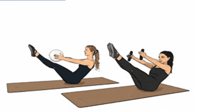
Swimming and stationary cycling with light resistance are often tolerated better than walking or impact activities. Always confirm with your treating clinician before starting.
Crutches are not a sign that you are failing to cope. They are the treatment. Taking weight off the hip is what allows the bone to recover.
Recovery and Returning to Normal Life
Recovery is measured in months, not weeks. Expect a gradual return rather than a sudden switch back to normal.
Most people can walk without aids within a few months, and full recovery is the usual outcome. Recurrence in the same hip is uncommon, though it can happen in the other hip.
- Follow your clinician’s plan for weaning off crutches rather than stopping suddenly.
- Rebuild strength slowly, starting with low-impact activity.
- Keep up vitamin D and calcium intake.
- Expect good days and setback days; that pattern is normal.
- Ask for repeat imaging if pain worsens instead of improving.
Working and Daily Life Adjustments
Desk work is usually manageable with a supportive chair and a footrest. Jobs that involve lifting, standing for long periods, or driving long distances may need temporary modification.
When to Seek Help Again
Contact your doctor if pain suddenly becomes much worse, if you develop fever, or if you cannot bear any weight at all. These signs suggest something other than a straightforward transient osteoporosis of the hip.
Also speak up if you are not improving after several months. A repeat MRI can confirm whether the edema is resolving and whether the diagnosis still fits.
Conclusion
Transient osteoporosis of the hip is painful, disruptive, and often frightening at the start, but it is a condition that usually resolves. The essential steps are an accurate MRI-based diagnosis, a period of protected weight bearing, sensible pain control, and a graded return to activity. With patience and the right plan, most people get back to their normal lives without surgery.
Frequently Asked Questions
Is transient osteoporosis of the hip permanent?
No. In most cases the bone changes are temporary and reverse over months. Bone density typically returns to normal, and the bone marrow edema disappears on follow-up imaging. A small number of people may have lingering discomfort, but permanent damage is uncommon.
How long does it usually last?
Many people notice significant improvement within three to six months, with full recovery often taking six to twelve months. Pregnancy-related cases sometimes settle faster, particularly in the weeks after delivery. Your own timeline depends on how strictly you protect the hip and how your body responds.
Is it the same as avascular necrosis?
They are different conditions, although early scans can look similar. Avascular necrosis involves death of bone tissue and carries a risk of joint collapse. Transient osteoporosis of the hip does not usually cause bone death and typically heals without surgery. This is exactly why MRI and specialist review matter.
Do I need surgery?
Most people do not. Surgery such as core decompression is reserved for unusual cases that fail to improve with conservative treatment or where the diagnosis is uncertain. The standard approach is crutches, physiotherapy, and time.
Can I keep walking on the affected leg?
Walking through the pain tends to make symptoms worse and can delay healing. Your doctor will usually recommend crutches or another walking aid during the painful phase. Short, protected steps around the house are generally fine, but avoid long walks or standing for extended periods.
Is it safe during pregnancy?
Yes, it is generally safe, and treatment focuses on rest, offloading, and gentle physiotherapy. Your care team will avoid medications and imaging that are not suitable during pregnancy. Symptoms often improve after delivery, and most women recover fully.
Which exercises are safe?
Non-weight-bearing activities such as swimming and light stationary cycling are usually well tolerated. Gentle range-of-motion exercises for the hip help prevent stiffness. Avoid running, jumping, heavy lifting, and deep squats until your clinician confirms the bone has recovered.
Will it come back?
Recurrence in the same hip is uncommon. Some people later develop the same problem in the opposite hip. Maintaining good vitamin D and calcium levels, avoiding prolonged immobilization, and addressing risk factors may lower the chance of a repeat episode.
Do I need supplements?
Only if blood tests show a deficiency or your doctor recommends them. Vitamin D and calcium are commonly checked because correcting low levels supports bone healing. Do not start high doses on your own, especially if you take other medications.
When can I return to sport?
Return to sport is usually guided by pain, strength, and follow-up imaging rather than a fixed date. Most people can build back toward impact activity over several months once they are pain-free and walking normally. A physiotherapist can set a safe progression for your sport.