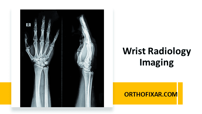
Wrist radiology imaging is a fundamental diagnostic tool in orthopedic and emergency medicine. The complex anatomy of the wrist, comprising eight carpal bones, the distal radius and ulna, and numerous articulations, requires systematic radiographic evaluation to identify pathology accurately. A thorough understanding of standard wrist radiographic views and their clinical applications is essential for medical practitioners.
Standard Wrist Radiology Views
The standard wrist radiology series consists of three essential views:
- Anteroposterior (AP) view
- Lateral view
- Scaphoid view
These three views provide comprehensive visualization of wrist anatomy and are sufficient for most clinical evaluations. Additional specialized views may be obtained based on clinical suspicion and specific diagnostic requirements.
Wrist Anteroposterior View (PA/AP View)
The wrist anteroposterior view serves as the cornerstone of wrist imaging evaluation. This projection provides optimal visualization of:
Anatomical Assessment:
- Bone shape and position evaluation
- Joint space analysis
- Bone density assessment
- Fracture detection and displacement evaluation
See Also: Wrist Anatomy: Bones, Ligaments & Joints

Key Radiographic Landmarks:
Gilula’s Lines (Carpal Arcs): These three smooth arcs represent the normal anatomical alignment of carpal bones:
- Arc 1: Proximal margins of scaphoid, lunate, and triquetrum
- Arc 2: Distal margins of scaphoid, lunate, and triquetrum
- Arc 3: Proximal margins of capitate and hamate
Disruption of these arcs suggests carpal instability or fracture-dislocation patterns.

Ulnar Variance Measurement: True ulnar variance is determined by:
- Drawing a line through the anterior cortex of the distal radius
- Creating a perpendicular line through the central reference point
- Measuring the distance between this line and the distal cortical rim of the ulnar dome
See Also: Ulnar Variance Measurement
Pathological Findings:
Scapholunate Dissociation:
- Normal scapholunate gap: approximately 2mm
- Terry Thomas sign: widening >4mm indicates dissociation
- Best visualized in ulnar deviation
Avascular Necrosis:
- Scaphoid: post-fracture complication
- Lunate: Kienböck disease
- Radiographic appearance: increased density, rarefaction, sclerotic changes
Triangular Fibrocartilage Complex (TFCC): May be visible on high-quality AP radiographs, though MRI remains the gold standard for TFCC evaluation.
Wrist Lateral View
The wrist lateral projection provides crucial information about:
Spatial Relationships:
- Scaphoid-lunate-radius alignment
- Metacarpal positioning relative to carpus
- Detection of carpal collapse patterns
Pathological Assessment:
- Fracture identification and displacement
- Soft tissue swelling around carpal bones
- Dorsal or volar angulation of fracture fragments
Angular Measurements: Critical for assessing carpal alignment and detecting subtle instability patterns.

Scaphoid View
This specialized oblique projection optimizes scaphoid visualization by:
- Elongating the scaphoid bone profile
- Reducing overlap with adjacent carpal bones
- Enhancing fracture line visibility
Clinical Significance: Scaphoid fractures are often occult on standard views, making this projection essential for suspected scaphoid injuries, particularly in young athletes with radial-sided wrist pain following fall on outstretched hand (FOOSH) injuries.
Specialized Radiographic Views
Carpal Tunnel View (Wrist Axial View)
Technique: Tangential projection of the carpal tunnel Clinical Applications:
- Hook of hamate fractures
- Trapezium fractures
- Carpal tunnel space assessment
- Pisiform positioning evaluation

Clenched-Fist AP View
Technique: AP projection obtained with patient making a fist Clinical Purpose:
- Dynamic assessment of carpal stability
- Detection of scapholunate dissociation
- Evaluation of ligamentous integrity under stress

Motion Views
Indications:
- Suspected carpal instability
- Dynamic scapholunate assessment
- Midcarpal joint evaluation
Types:
- Radial deviation view
- Ulnar deviation view
- Flexion and extension views
Clinical Applications
Skeletal Age Assessment
The left wrist and hand are standardly used for skeletal age determination due to:
- Multiple ossification centers
- Systematic ossification progression
- Reduced environmental influence on non-dominant hand
Methodology:
- Comparison with standardized reference plates
- Gender-specific standards
- Assessment of carpal bones, metacarpal epiphyses, and phalangeal growth plates
Normal Variants:
- Two-thirds of population within ±1 year of chronological age
- ≥3 years variance considered abnormal
Emergency Department Applications
Fracture Detection:
- Distal radius fractures (most common wrist fracture)
- Scaphoid fractures (most common carpal fracture)
- Ulnar styloid fractures
- Carpal fractures and dislocations
Instability Patterns:
- Scapholunate dissociation
- Lunotriquetral dissociation
- Midcarpal instability
- Distal radioulnar joint (DRUJ) instability
Advanced Imaging Considerations
While conventional radiography remains the first-line imaging modality, certain clinical scenarios require advanced imaging:
MRI Indications:
- TFCC pathology
- Occult fractures
- Soft tissue injury assessment
- Avascular necrosis staging
CT Applications:
- Complex fracture evaluation
- Preoperative planning
- Hardware assessment
- Arthritis progression monitoring
Conclusion
Wrist radiology imaging requires systematic approach and thorough understanding of normal anatomy and common pathological patterns. The combination of standard three-view series with selective use of specialized projections provides comprehensive diagnostic capability for most wrist disorders. Recognition of key radiographic landmarks, measurement techniques, and pathological patterns enables accurate diagnosis and appropriate treatment planning.
Continuous correlation with clinical findings and judicious use of advanced imaging modalities when indicated ensures optimal patient care and diagnostic accuracy in wrist pathology evaluation.

References & More
- chuind FA, Linscheid RL, An KN, et al. A normal database of posteroanterior roentgenographic measurements of the wrist. J Bone Joint Surg Am. 1992;74:1418–1429. PubMed
- Feipel V, Rinnen D, Rooze M. Postero-anterior radiography of the wrist: normal database of carpal measurements. Surg Radiol Anat. 1998;20(3):221–226. PubMed
- Medoff RJ. Essential radiographic evaluation for distal radius fractures. Hand Clin. 2005;21(3):279–288. PubMed
- De Filippo M, Sudberry JJ, Lombardo E, Corradi M, Pogliacom F, Ferrari FS, Bocchi C, Zompatori M. Pathogenesis and evolution of carpal instability: imaging and topography. Acta Biomed. 2006 Dec;77(3):168-80. PMID: 17312988. PubMed
- Hansman CF, Mresh MM. Appearance and fusion of ossification centers in the human skeleton. Am J Roentgenol. 1962;88:476–482. PubMed
- Orthopedic Physical Assessment by David J. Magee, 7th Edition.


