Approach
Volar Approach to the Wrist

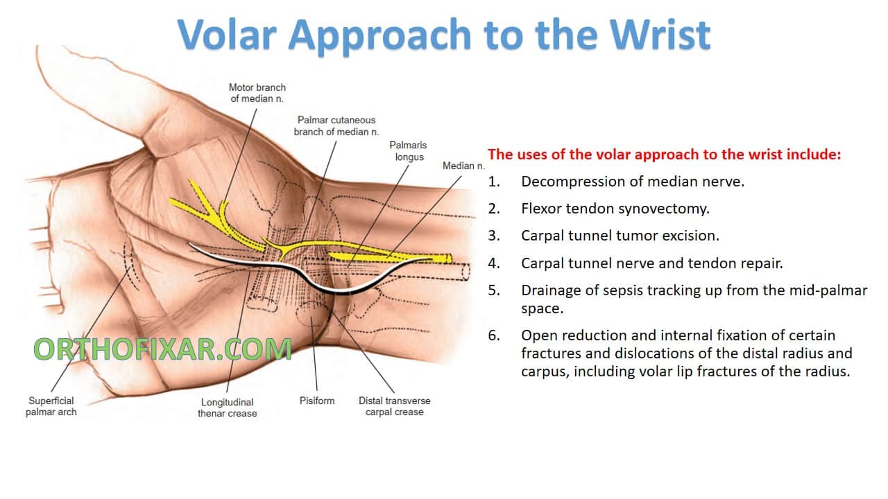
The most common use of the Volar Approach to the Wrist is Decompression of median nerve (carpal tunnel syndrome).
The other uses of the volar approach to the wrist include:
- Flexor tendon synovectomy.
- Carpal tunnel tumor excision.
- Carpal tunnel nerve and tendon repair.
- Drainage of sepsis tracking up from the mid-palmar space.
- Open reduction and internal fixation of certain fractures and dislocations of the distal radius and carpus, including volar lip fractures of the radius.
See Also: Carpal Tunnel Syndrome
Position of the Patient
- Place the patient supine on an operating table.
- Rest the forearm on a hand table in the supinated position so that the palm faces upward. Use an exsanguinating bandage.

Landmarks and Incision
- Landmark:
- Thenar crease.
- Transverse skin crease .
- Tendon of the palmaris longus muscle .
- Incision:
- Begin the incision just to the ulnar side of the thenar crease, about one third of the way into the hand.
- Curve it proximally, remaining just to the ulnar side of the thenar crease, until the flexion crease of the wrist is almost reached: to avoid problems in skin healing, do not wander into the thenar crease itself.
- Then, curve the incision toward the ulnar side of the forearm so that the flexion crease is not crossed transversely

Internervous plane
- There is No true internervous plane for the volar approach to the wrist.
- No muscles are transected:
- Abductor pollicis brevis and palmaris brevis fibers that cross the midline can occasionally be dissected.
- True anatomic dissection:
- Major nerves identified, dissected out and preserved.
- Plane of dissection between median nerve and flexor carpi radialis tendon.
Superficial dissection
- Incise skin flaps then incise fat.
- Section fibers of superficial palmar fascia in line with incision.
- Retract curved flaps medially to expose insertion of palmaris longus into flexor retinaculum,
- Retract palmaris longus tendon medially to expose median nerve between palmaris longus and flexor carpi radialis tendon,
- Pass a blunt, flat instrument (such as a McDonald dissector) down the carpal tunnel between the flexor retinaculum and the median nerve.
- Incise entire length of retinaculum / transverse carpal ligament on ulnar side of the nerve.


Deep dissection
- Identify motor branch of the median nerve (anterolateral side of the median nerve as it emerges from carpal tunnel).
- If require access to volar aspect of wrist joint:
- Mobilize median nerve and retract radially (so you don’t stretch motor branch).
- Mobilize and retract flexor tendons.
- Incise base of carpal tunnel longitudinally.
- The most convenient approach for access to the volar aspect of the distal radius is the distal portion of the volar approach to the radius .


Approach Extension
Proximal Extension:
The volar approach to the wrist can be extended to expose the median nerve, to accomplish this:
- extend the skin incision proximally, running it up the middle of the anterior surface of the forearm.
- Incise the deep fascia of the forearm between the palmaris longus and flexor carpi radialis muscles.
- Retract the flexor carpi radialis in a radial direction and the palmaris longus in an ulnar direction, exposing the muscle belly of the flexor digitorum superficialis muscle in the distal two thirds of the forearm.
- The median nerve adheres to the deep surface of the flexor digitorum superficialis, held there by fascia.
- Thus, if the flexor digitorum superficialis is reflected, the nerve goes with it.
Distal Extension:
- The volar approach to the wrist can be extended into a volar zigzag approach for any of the fingers, providing complete exposure of all the palmar structures (Volar Approach to the Flexor Tendons).

Dangers
The structures at risk during the volar approach to the wrist include:
- Palmar cutaneous branch of median nerve:
- Arises 5 cm proximal to wrist joint.
- Runs ulnar to flexor carpi radialis tendon before crossing flexor retinaculum.
- Greatest risk if the skin incision is not angled to the ulnar side of the forearm .
- Motor branch of median nerve:
- Significant anatomic variation.
- Risk to nerve minimized if incision through retinaculum made ulnar to median nerve.
- Superficial palmar arch:
- Crosses palm at level of distal end of outstretched thumb.
- In danger if flexor retinaculum blindly cut (can go too far distally)
- Avoid injury if retinaculum is cut under direct observation for its entire length.
References
- Surgical Exposures in Orthopaedics book – 4th Edition
- Campbel’s Operative Orthopaedics book 12th
Last Reviewed
June 11, 2023
June 11, 2023
Contributed by
OrthoFixar
OrthoFixar
Orthofixar does not endorse any treatments, procedures, products, or physicians referenced herein. This information is provided as an educational service and is not intended to serve as medical advice.
Angle Meter App for Android & iOS
- Lifetime product updates
- Install on one device
- Lifetime product support
Orthopedic FRCS VIVAs Quiz
- Lifetime product updates
- Install on one device
- Lifetime product support
Top 12 Best Free Orthopedic Apps
- Lifetime product updates
- Install on one device
- Lifetime product support
All-in-one Orthopedic App
- Lifetime product updates
- Install on one device
- Lifetime product support