Cleidocranial dysplasia (CCD), also known as cleidocranial dysostosis, is a rare inherited skeletal disorder characterized by abnormal development of the clavicles, skull, teeth, and other skeletal structures. It is most commonly caused by a pathogenic variant in the RUNX2 gene, which plays an essential role in osteoblast differentiation and bone formation.
The disorder primarily affects bones formed through intramembranous ossification, particularly the clavicles and cranial bones. However, skeletal structures developing through endochondral ossification can also be involved.
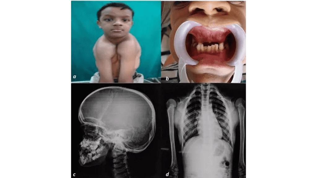
Characteristic findings include absent or underdeveloped clavicles, delayed closure of cranial sutures and fontanelles, short stature, hypermobile shoulders, and delayed eruption of permanent teeth.
Although many individuals experience relatively mild skeletal limitations, significant dental abnormalities and orthopedic complications may require specialized treatment.
This article provides an evidence-based overview of the genetics, pathophysiology, clinical presentation, radiological features, diagnosis, and management of cleidocranial dysplasia.
What Is Cleidocranial Dysplasia?
Cleidocranial dysplasia is a genetic skeletal dysplasia affecting bone growth, development, and maintenance.
The term cleidocranial refers to two of the most frequently affected skeletal structures:
- Cleido: Clavicle or collarbone.
- Cranial: Skull and cranial bones.
- Dysplasia: Abnormal development of tissues or structures.
The disorder displays considerable clinical variability. Some individuals have the classic cranial, clavicular, and dental abnormalities, whereas others exhibit only mild skeletal changes or isolated dental manifestations.
The Classic Triad of Cleidocranial Dysplasia
Three clinical findings are especially characteristic:
- Hypoplastic or absent clavicles, causing increased shoulder mobility.
- Delayed closure of cranial sutures and fontanelles, sometimes persisting into adulthood.
- Dental abnormalities, including delayed eruption of permanent teeth, retained primary teeth, and supernumerary teeth.
Recognition of this triad is important for early diagnosis.
Clinical pearl: A patient who can bring both shoulders unusually close together in front of the chest, especially when associated with dental abnormalities or persistent open fontanelles, should be evaluated for cleidocranial dysplasia.
Causes and Genetics of Cleidocranial Dysplasia
RUNX2 Gene Mutation
The principal genetic cause of cleidocranial dysplasia is a heterozygous pathogenic variant in the RUNX2 gene, located on chromosome 6p21.1.
RUNX2 encodes a transcription factor essential for the development and differentiation of osteoblasts, the cells responsible for forming bone.
Under normal circumstances, RUNX2 regulates genes involved in skeletal development and bone formation.
When RUNX2 function is impaired, osteoblast differentiation and skeletal development are disrupted, producing the characteristic cranial, clavicular, dental, and other skeletal abnormalities.
Role of RUNX2 in Bone Formation
Bone develops through two major mechanisms.
Intramembranous Ossification
Intramembranous ossification involves the direct differentiation of mesenchymal cells into osteoblasts without an intermediate cartilage model.
It contributes to the formation of several bones, including:
- Flat bones of the skull.
- Much of the clavicles.
- Portions of the facial skeleton.
These structures are prominently affected in cleidocranial dysplasia.
Endochondral Ossification
Endochondral ossification involves the replacement of a cartilage model with bone.
Although cleidocranial dysplasia preferentially affects intramembranous bone formation, endochondral skeletal structures may also exhibit abnormalities.
This explains findings such as coxa vara, short stature, pelvic abnormalities, and shortened phalanges.
Inheritance Pattern
Cleidocranial dysplasia is typically inherited in an autosomal dominant pattern.
This means that a pathogenic variant affecting one copy of RUNX2 is generally sufficient to cause the condition.
An affected individual has a 50% chance of transmitting the pathogenic variant to each child.
However, some individuals develop the disorder because of a new (de novo) genetic variant, without a previously affected parent.
Clinical severity can vary considerably, even among members of the same family.
A minority of individuals with a CCD-like phenotype may have other molecular causes, including pathogenic variants involving CBFB.
Clinical Features of Cleidocranial Dysplasia
The manifestations of cleidocranial dysplasia involve multiple regions of the skeleton and the dentition.
1. Clavicular Abnormalities
The most distinctive skeletal abnormality is hypoplasia or aplasia of the clavicles.
Affected patients may demonstrate:
- Partial or complete absence of one or both clavicles.
- Narrow, sloping shoulders.
- Excessive shoulder mobility.
- Ability to approximate the shoulders anteriorly.
- Abnormal shoulder contour.
The degree of clavicular involvement varies. Some individuals retain partially developed clavicles, whereas others have marked bilateral absence.
Despite these anatomical abnormalities, shoulder function can remain remarkably well preserved.
Consequently, many patients do not require surgical treatment of the clavicles.
2. Cranial and Facial Abnormalities
Skull involvement is another major feature of cleidocranial dysplasia.
Typical manifestations include:
- Delayed closure of cranial sutures.
- Persistently open anterior fontanelle.
- Frontal bossing.
- Parietal bossing.
- Broad forehead.
- Midfacial hypoplasia.
- Depressed nasal bridge.
- Hypertelorism.
- Abnormal skull shape.
Wormian Bones
Wormian bones are accessory ossicles located within cranial sutures.
They may be observed in cleidocranial dysplasia, often around the lambdoid sutures.
However, Wormian bones are not specific to this condition and can also occur in other skeletal disorders and in individuals without skeletal disease.
Delayed Fontanelle Closure
The anterior fontanelle may remain open considerably longer than expected and may persist into adulthood.
In patients with substantial cranial vault defects, additional precautions may be necessary to prevent traumatic injury.
3. Dental Abnormalities
Dental manifestations are among the most clinically important features of cleidocranial dysplasia.
Common abnormalities include:
- Delayed eruption of permanent teeth.
- Retention of primary or deciduous teeth.
- Supernumerary teeth.
- Impacted permanent teeth.
- Dental crowding.
- Malocclusion.
- Abnormal tooth positioning.
Some individuals have numerous unerupted permanent teeth that require coordinated surgical and orthodontic treatment.
These dental manifestations can affect mastication, facial appearance, oral hygiene, speech, and quality of life.
Important: Early referral to a dental team experienced in cleidocranial dysplasia can substantially improve long-term treatment planning.
4. Short Stature and Growth Abnormalities
Individuals with cleidocranial dysplasia frequently have mild to moderate short stature.
Short stature is usually relatively proportionate, although associated skeletal abnormalities may alter body proportions.
Growth abnormalities reflect disturbances in normal skeletal development.
Intellectual development is typically normal.
5. Pelvic and Hip Abnormalities
The pelvis and proximal femur may be affected.
Recognized findings include:
- Delayed ossification of the pubic bones.
- Abnormal development of the pubic symphysis.
- Narrow or otherwise abnormal pelvic morphology.
- Coxa vara.
- Hip-related mechanical abnormalities.
Coxa Vara in Cleidocranial Dysplasia
Coxa vara is a deformity characterized by a reduced femoral neck–shaft angle.
When pronounced, it may cause:
- Limping or gait abnormalities.
- Hip pain.
- Restricted hip movement.
- Abnormal lower-limb biomechanics.
- Functional limitations.
Mild cases may require only monitoring. Severe or progressive deformity may require corrective surgery.
6. Hand and Finger Abnormalities
The hands may demonstrate characteristic skeletal changes.
These include:
- Brachydactyly.
- Shortened middle phalanges.
- Hypoplastic distal phalanges.
- Short, broad thumbs.
- Cone-shaped epiphyses.
- Pseudoepiphyses of the metacarpals.
Shortening and deformity of the middle phalanges may be particularly evident in the third, fourth, and fifth fingers.
7. Other Associated Manifestations
Additional findings may include:
- Scoliosis.
- Genu valgum.
- Pes planus.
- Recurrent shoulder or elbow dislocation.
- Reduced bone mineral density.
- Increased susceptibility to fractures in some patients.
- Recurrent middle ear infections.
- Conductive hearing loss.
- Recurrent sinus infections.
- Upper airway obstruction or sleep-disordered breathing.
The presence and severity of these abnormalities vary between patients.
Radiographic Findings of Cleidocranial Dysplasia
Radiological assessment plays a central role in diagnosing cleidocranial dysplasia.
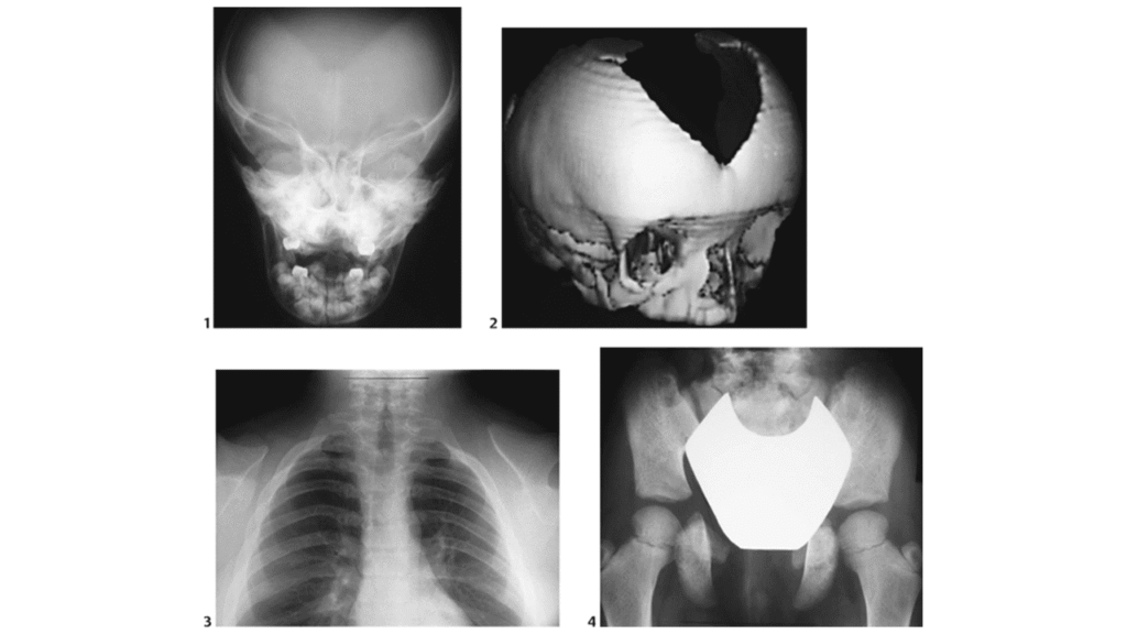
A skeletal survey may demonstrate characteristic abnormalities involving the clavicles, skull, pelvis, hips, hands, and feet.
Skull X-Ray Findings
Typical findings include:
- Delayed closure of cranial sutures.
- Persistently open fontanelles.
- Wormian bones.
- Frontal and parietal bossing.
- Abnormal cranial ossification.
These changes support the diagnosis when accompanied by the characteristic clavicular and dental findings.
Chest X-Ray Findings
A chest radiograph may demonstrate:
- Complete or partial absence of the clavicles.
- Hypoplastic clavicles.
- Abnormal clavicular morphology.
- A narrow, sloping shoulder configuration.
Radiographic hallmark: Hypoplastic or absent clavicles are highly characteristic, particularly when associated with delayed cranial suture closure.
Pelvic X-Ray Findings
Pelvic radiographs may show:
- Delayed ossification of the pubic bones.
- Abnormal pubic symphysis.
- Pelvic skeletal hypoplasia.
- Coxa vara.
- Abnormalities involving the iliac bones.
Hand X-Ray Findings
Hand radiographs may reveal:
- Short middle phalanges.
- Hypoplastic distal phalanges.
- Cone-shaped epiphyses.
- Pseudoepiphyses involving the metacarpals.
Dental Radiographic Findings
Panoramic dental radiography may show:
- Multiple unerupted permanent teeth.
- Supernumerary teeth.
- Retained primary teeth.
- Abnormal tooth positioning and impaction.
Additional dental imaging may be required for complex orthodontic and surgical planning.
Summary of Important Radiographic Findings
| Anatomical region | Characteristic findings |
|---|---|
| Skull | Delayed suture closure, open fontanelles, Wormian bones |
| Clavicles | Hypoplasia or aplasia |
| Pelvis | Delayed pubic ossification, abnormal pubic symphysis |
| Hip | Coxa vara |
| Hands | Shortened phalanges, cone-shaped epiphyses, pseudoepiphyses |
| Teeth | Supernumerary teeth, impaction, delayed eruption |
Diagnosis of Cleidocranial Dysplasia
Diagnosis is based on the combination of clinical findings, skeletal imaging, dental assessment, and, when appropriate, molecular genetic testing.
Clinical Examination
A clinician should assess:
- Shoulder position and mobility.
- Presence or absence of palpable clavicles.
- Cranial shape and fontanelle closure.
- Dental development.
- Growth parameters.
- Hip movement and gait.
- Hand and finger morphology.
- Family history.
Radiological Evaluation
Appropriate radiographs help identify characteristic skeletal abnormalities.
Depending on clinical findings, investigations may include skull, chest, pelvic, hand, and dental imaging.
Genetic Testing
Molecular testing can identify a pathogenic or likely pathogenic variant in RUNX2.
Testing may involve:
- RUNX2 sequence analysis.
- RUNX2 deletion and duplication analysis.
- A broader skeletal dysplasia gene panel when indicated.
A negative RUNX2 result does not automatically exclude a CCD-spectrum disorder. Additional genetic evaluation may be necessary when clinical suspicion remains high.
Genetic counseling should be offered to affected individuals and families.
Differential Diagnosis
Several disorders may share individual clinical or radiographic features with cleidocranial dysplasia.
Important considerations include:
- Pycnodysostosis: May cause short stature, open cranial sutures, and dental abnormalities, typically with increased bone density.
- Hypophosphatasia: A mineralization disorder that can produce skeletal abnormalities and premature tooth loss.
- Osteogenesis imperfecta: May produce Wormian bones and skeletal abnormalities, often accompanied by bone fragility.
- Mandibuloacral dysplasia: Can involve clavicular hypoplasia and characteristic craniofacial and skeletal changes.
- CBFB-related cleidocranial dysplasia: May closely resemble RUNX2-related disease.
Clinical assessment and appropriately selected laboratory, radiological, and genetic investigations help distinguish these disorders.
Treatment and Management of Cleidocranial Dysplasia
There is currently no established treatment that reverses the underlying genetic abnormality.
Management is individualized and focuses on improving function, preventing complications, and addressing dental, orthopedic, and other medical needs.
A multidisciplinary approach is often beneficial.
1. Management of Clavicular Abnormalities
Most patients with hypoplastic or absent clavicles do not require surgical intervention.
Because shoulder function is often well preserved, treatment is generally conservative.
Management may include:
- Clinical observation.
- Assessment of shoulder function.
- Physical therapy when indicated.
- Counseling regarding the appearance of the shoulders.
Surgical reconstruction or lengthening may be considered in selected cases, particularly for significant cosmetic or functional concerns, but it is not routinely indicated.
2. Treatment of Coxa Vara
Management depends on the severity of deformity, symptoms, and functional impairment.
Mild, stable deformities may be monitored.
Patients with significant pain, progressive deformity, or gait disturbance should undergo orthopedic evaluation.
Corrective Osteotomy
A severe or symptomatic coxa vara deformity may require a proximal femoral valgus osteotomy.
This operation aims to improve the femoral neck–shaft alignment and hip biomechanics.
Surgical planning depends on individual anatomy, deformity severity, and the patient’s clinical needs.
Internal fixation is typically used to maintain the corrected position while bone healing occurs.
Not every patient with coxa vara requires surgery.
3. Dental and Orthodontic Treatment
Dental treatment is frequently one of the most important aspects of long-term care.
A coordinated approach involving pediatric dentists, orthodontists, and oral and maxillofacial surgeons may be required.
Management options include:
- Extraction of selected retained primary teeth.
- Removal of supernumerary teeth.
- Surgical exposure of impacted permanent teeth.
- Orthodontic traction and alignment.
- Prosthetic dental rehabilitation when necessary.
Dental treatment often requires multiple stages and should be planned according to the patient’s age, dental development, and specific anatomical findings.
Simply waiting indefinitely for impacted permanent teeth to erupt is generally not an effective management strategy.
4. Management of Cranial Abnormalities
Most patients do not require cranial surgery.
However, individuals with large persistent cranial defects may need:
- Evaluation by craniofacial specialists.
- Protection against head injury.
- Protective helmets during activities with increased trauma risk.
- Selected surgical procedures when clinically appropriate.
5. Bone Health and Orthopedic Monitoring
Patients may require periodic evaluation for:
- Scoliosis.
- Genu valgum.
- Pes planus.
- Hip deformities.
- Recurrent joint dislocations.
- Reduced bone mineral density.
Bone mineral density assessment may be appropriate from adolescence, with subsequent monitoring individualized according to clinical findings.
Calcium and vitamin D intake should be assessed, with supplementation when clinically indicated.
6. Hearing and Respiratory Management
Recurrent middle ear infections and conductive hearing loss are recognized complications.
Audiological assessment and otolaryngology referral should be considered when indicated.
Sleep assessment may be required for symptoms suggesting upper airway obstruction or obstructive sleep apnea.
7. Long-Term Follow-Up
Regular follow-up should be individualized and may involve:
- Orthopedic specialists.
- Pediatric or adult dental specialists.
- Orthodontists.
- Clinical geneticists.
- Audiologists.
- Otolaryngologists.
- Craniofacial specialists.
The overall goal is to maintain function, protect long-term skeletal and dental health, and improve quality of life.
Prognosis and Life Expectancy
The prognosis for individuals with cleidocranial dysplasia is generally favorable.
Many affected individuals lead active, independent lives with relatively minor skeletal limitations.
Intellectual development is typically normal.
The absence or hypoplasia of clavicles often has limited impact on everyday function.
However, dental abnormalities may require substantial treatment, and some patients develop orthopedic, hearing, respiratory, or bone-health complications.
There is no established characteristic reduction in life expectancy for individuals with uncomplicated cleidocranial dysplasia.
Early recognition, appropriate monitoring, and access to multidisciplinary care can help reduce preventable complications.
Cleidocranial Dysplasia: Key Points for Medical Students
- Cleidocranial dysplasia is a predominantly autosomal dominant skeletal disorder.
- It is most commonly caused by pathogenic variants in RUNX2.
- RUNX2 is a transcription factor involved in osteoblast differentiation.
- The disorder prominently affects bones formed by intramembranous ossification.
- The classic triad includes clavicular hypoplasia or aplasia, delayed cranial suture closure, and dental abnormalities.
- Shoulder hypermobility is an important clinical clue.
- Wormian bones may be seen on skull radiographs.
- Coxa vara and shortening of the phalanges are recognized skeletal findings.
- Most clavicular abnormalities do not require surgery.
- Severe symptomatic coxa vara may require corrective osteotomy.
- Specialist dental and orthodontic management is frequently necessary.
- Normal intelligence and a generally favorable long-term outlook are typical.
Frequently Asked Questions About Cleidocranial Dysplasia
What is the main cause of cleidocranial dysplasia?
Cleidocranial dysplasia is most commonly caused by a pathogenic variant in the RUNX2 gene, which regulates osteoblast differentiation and skeletal development.
Is cleidocranial dysplasia inherited?
Yes. It is typically inherited in an autosomal dominant pattern, although new genetic variants can arise in individuals without a family history.
What are the three classic features of cleidocranial dysplasia?
The three classic features are hypoplastic or absent clavicles, delayed closure of cranial sutures and fontanelles, and characteristic dental abnormalities.
Why are the clavicles absent in cleidocranial dysplasia?
Impaired RUNX2 function disrupts normal bone development, especially intramembranous ossification, contributing to incomplete development or absence of the clavicles.
What are Wormian bones?
Wormian bones are accessory bones that develop within cranial sutures. They may be seen in cleidocranial dysplasia but are not specific to the disorder.
What dental problems occur in cleidocranial dysplasia?
Common dental problems include delayed eruption of permanent teeth, supernumerary teeth, retained primary teeth, impacted teeth, and malocclusion.
Can cleidocranial dysplasia be cured?
There is currently no established cure for the underlying genetic disorder. However, dental, orthopedic, and other associated complications can often be managed effectively.
Does cleidocranial dysplasia affect intelligence?
Intelligence is typically normal in individuals with cleidocranial dysplasia.
Is surgery required for absent clavicles?
Usually not. Many patients have satisfactory shoulder function despite clavicular hypoplasia or absence. Surgery may be considered in selected cases.
How is coxa vara treated in patients with cleidocranial dysplasia?
Mild cases may be monitored, while severe, progressive, or symptomatic deformities may require corrective proximal femoral valgus osteotomy.
Conclusion
Cleidocranial dysplasia is a rare genetic skeletal disorder caused predominantly by pathogenic variants in RUNX2, resulting in abnormal bone development and characteristic abnormalities of the clavicles, skull, and teeth.
The most important diagnostic features are hypoplastic or absent clavicles, delayed closure of cranial sutures, and dental abnormalities.
Additional manifestations include short stature, shoulder hypermobility, Wormian bones, coxa vara, pelvic ossification abnormalities, and shortening of the phalanges.
Diagnosis relies on clinical and radiographic evaluation, supported by molecular genetic testing when appropriate.
Most clavicular abnormalities require no surgical treatment, but patients with significant orthopedic deformity or dental complications may benefit from specialized interventions.
Early diagnosis and multidisciplinary management are essential for optimizing long-term functional and oral health outcomes.
References and Trusted Medical Sources
- Machol K, Mendoza-Londono R, Lee B. Cleidocranial Dysplasia Spectrum Disorder. GeneReviews®. University of Washington, Seattle. Updated April 13, 2023. NCBI Bookshelf. PMID: 20301686.
- Farrow E, Nicot R, Wiss A, Laborde A, Ferri J. Cleidocranial Dysplasia: A Review of Clinical, Radiological, Genetic Implications and a Guidelines Proposal. Journal of Craniofacial Surgery. 2018;29(2):382–389. PubMed. DOI: 10.1097/SCS.0000000000004200.
- The Impact of RUNX2 Gene Variants on Cleidocranial Dysplasia Phenotype: A Systematic Review. PubMed Central – Full Text.
- Diagnosis, Treatment, and Management Recommendations for Cleidocranial Dysplasia: A Modified Delphi Panel. PubMed Central – Full Text.