Orthopedic care depends heavily on imaging. A patient may begin with an X-ray after an injury, undergo an MRI for suspected ligament damage, receive a CT scan for surgical planning, and return for follow-up images during recovery. The challenge is no longer simply producing high-quality images. It is making sure those images, reports, measurements, and prior comparisons are available to the right clinician at the right stage of care.
That challenge has made connected imaging one of the most important digital health trends in musculoskeletal medicine. In January 2026, the Office of the National Coordinator for Health Information Technology highlighted continuing difficulties with the access, exchange, and use of diagnostic images across healthcare settings. The agency’s request for information reflects a broader shift: medical images are increasingly being treated as essential parts of the longitudinal health record, not isolated files inside a radiology department.
Why Orthopedic Imaging Is Becoming More Connected
Musculoskeletal care rarely occurs within a single department. An emergency physician may order the first radiograph, a radiologist may interpret it, an orthopedic surgeon may review the study before deciding on treatment, and a rehabilitation specialist may later need the report to understand restrictions.
When these professionals work in separate systems, important context can be lost. Images may arrive on discs, links may expire, prior studies may be unavailable, or reports may be copied into the chart without access to the underlying images. Clinicians can then spend time searching for records, requesting transfers, or repeating studies completed elsewhere.
Connected imaging addresses this problem by linking image storage, diagnostic viewing, reporting, and the electronic health record.
What PACS Medical Imaging Software Adds to Orthopedic Care
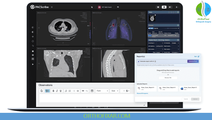
PACS medical imaging software provides the digital infrastructure used to store, retrieve, display, and share studies from modalities such as X-ray, CT, MRI, and ultrasound. DICOM remains the principal standard that allows imaging devices, archives, and viewing systems to exchange medical image data. DICOMweb extends that framework through web-based services for searching, retrieving, and storing DICOM objects.
In orthopedics, the value of PACS goes beyond storage. Clinicians often need to compare images across multiple dates, inspect alignment, review fracture healing, measure anatomical structures, and collaborate with colleagues. A modern viewer lets authorized users open studies across approved devices.
This is useful for trauma referrals, multidisciplinary reviews, postoperative follow-ups, and second opinions. A surgeon receiving a transferred patient can review the original images before arrival. A radiologist can compare current findings with earlier studies. A clinician at another location can examine the same series while discussing the case with the treating team.
The result is better continuity between diagnosis, treatment planning, intervention, and recovery monitoring.
Cloud Access Is Changing Where Images Can Be Reviewed
Traditional imaging systems were commonly built around local servers and dedicated workstations. These environments still meet many hospital needs, but orthopedic care now extends across outpatient clinics, urgent care centers, imaging facilities, ambulatory surgery centers, and remote consultation settings.
Cloud-based access supports this distributed model. Images can be made available to authorized clinicians across locations while centralizing storage and administration. Web-based viewing may reduce dependence on a specific workstation, which is valuable when surgeons divide their time between clinics, hospitals, and operating facilities.
However, cloud deployment is not a simple hosting decision. Healthcare organizations must evaluate encryption, identity management, role-based permissions, audit trails, backups, availability, data residency, and disaster recovery. Clinical performance also matters. Large CT and MRI studies must load reliably, and image quality must remain appropriate for the intended diagnostic use.
The Impact on Common Orthopedic Workflows
Trauma and Emergency Referrals
Fracture care often involves transfers between facilities. Immediate access to original radiographs and CT images can help the receiving team understand fracture configuration, evaluate displacement, and prepare equipment or specialist coverage before arrival.
Preoperative Planning
Surgeons may use radiographs, CT, or MRI to evaluate anatomy, alignment, bone loss, soft-tissue injury, and implant considerations. Keeping current and prior studies together reduces fragmentation during planning and makes multidisciplinary review easier.
Sports Medicine
Sports injuries frequently require comparison between clinical examination, X-ray, ultrasound, and MRI findings. Connected systems can help physicians and therapists work from the same diagnostic information while coordinating treatment and return-to-activity decisions.
Postoperative Follow-Up
Serial imaging is central to monitoring fracture union, implant position, alignment, and complications. Access to prior studies allows clinicians to identify meaningful change rather than evaluating each follow-up image in isolation.
Remote Consultation and Second Opinions
Orthopedic subspecialists are not available in every location. Secure image sharing can make specialist review practical for complex spine, trauma, pediatric, tumor, or joint reconstruction cases without requiring physical media.
Structured Reporting Is the Next Important Layer
Access to images solves only part of the problem. Orthopedic care also depends on consistent reporting of measurements, anatomical location, severity, alignment, implant status, and comparison with earlier studies.
Structured reporting can make these findings easier to review and reuse. Instead of leaving important measurements buried in free text, systems can associate them with the relevant study or image. This may support clearer longitudinal comparison, quality review, registry participation, and communication between radiologists and orthopedic teams.
Human review remains essential. New imaging technologies, including advanced decision-support models, should assist qualified clinicians rather than remove clinical oversight. A 2026 musculoskeletal imaging study reported progress in models that work across fracture detection, osteoarthritis grading, and bone tumor classification, while noting the challenge of generalizing across diseases and anatomical regions.
What Healthcare Organizations Should Evaluate
Orthopedic practices and health systems assessing an imaging platform should begin with workflow rather than feature volume. They should determine where studies originate, who needs access, which measurement and annotation tools are required, how reports are finalized, and where results must appear.
They should also examine DICOM conformance, viewer performance, EHR launch context, patient matching, auditability, access controls, backup procedures, and external image exchange. Integration testing should include real scenarios, such as transferred trauma cases, duplicate patient records, corrected reports, unavailable prior studies, and users working across multiple sites.
A system can be technically connected yet remain difficult to use. The goal should be to reduce navigation and manual handoffs without hiding information clinicians need to make safe decisions.
Connected Imaging Is Becoming Part of Orthopedic Infrastructure
The future of orthopedic imaging is not defined by one modality or software feature. It is defined by whether information can move with the patient.
X-rays, CT scans, MRIs, reports, measurements, and prior comparisons should remain connected as the patient progresses from diagnosis to treatment and recovery. PACS provides the imaging foundation, cloud access expands availability, and EHR integration places those studies within the wider clinical record.
As interoperability standards mature and healthcare organizations focus more closely on diagnostic image exchange, connected imaging will become less of an optional technology project and more of a basic requirement for coordinated musculoskeletal care. For orthopedic teams, the central question is no longer whether images are digital. It is whether those images are available, clinically usable, and connected when decisions must be made.