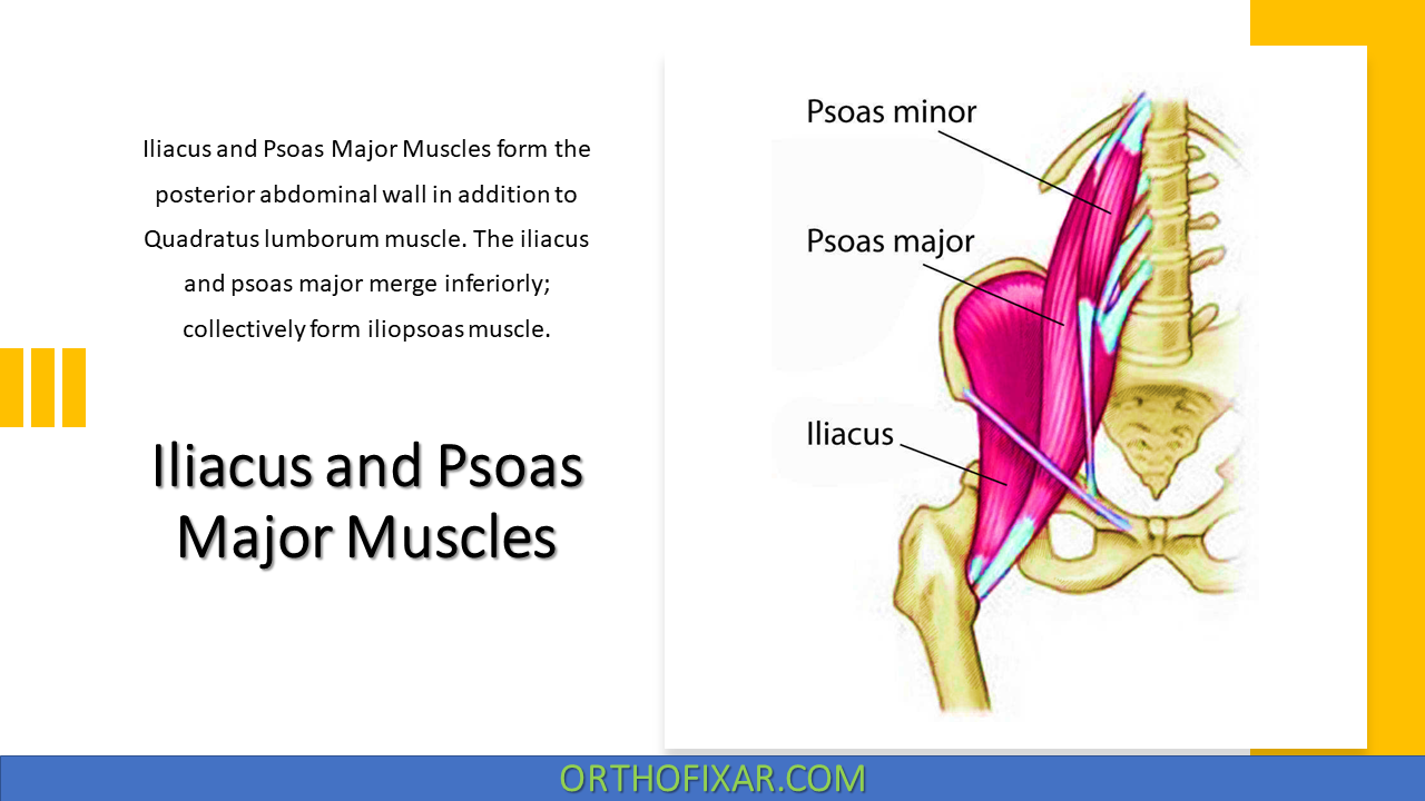
Iliacus and Psoas Major Muscles
Iliacus and Psoas Major Muscles are one of the posterior abdominal wall in addition to Quadratus lumborum muscle. The…
Read Full Entry →Clinical anatomy of the human body. Upper & lower limb, Head & neck, chest, abdomen and spine anatomy. Anatomy of bones, muscles, ligaments and joints.
Iliacus and Psoas Major Muscles are one of the posterior abdominal wall in addition to Quadratus lumborum muscle. The…
Read Full Entry →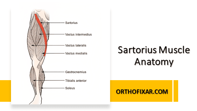
The sartorius muscle, the “tailor’s muscle” (L. sartus, patched or repaired), is long and ribbon-like. It passes lateral…
Read Full Entry →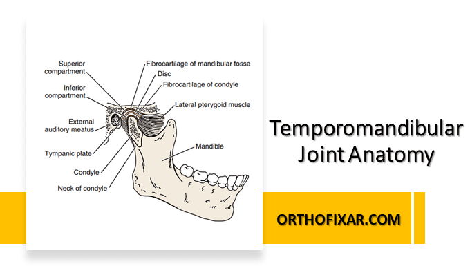
The temporomandibular joints (TMJs) are two of the most frequently used joints in the body, but they probably…
Read Full Entry →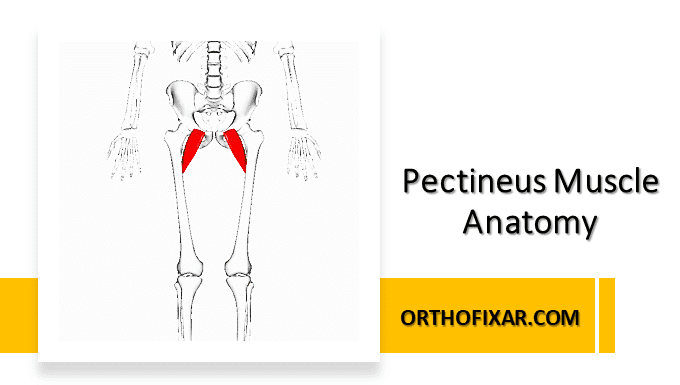
The pectineus muscle originates from the pectineal line (pecten pubis) and adjacent bone of pelvis, and inserts on…
Read Full Entry →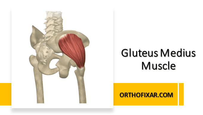
The Gluteus medius muscle is a fan-shaped appearance muscle that lies between the gluteus maximus and gluteus minimus…
Read Full Entry →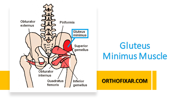
Both the gluteus medius and gluteus minimus, are fan shaped, and their fibers converge in the same manner…
Read Full Entry →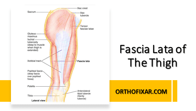
Fascia lata and Tensor fascia latae are different structures. The Fascia lata is a fibrous tissue that lies…
Read Full Entry →
The tensor fasciae lata muscle originates from the anterior superior iliac spine (ASIS) and anterior aspect of the…
Read Full Entry →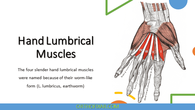
The 1st lumbrical muscle originates from the radial side of the most radial tendon of the flexor digitorum…
Read Full Entry →
The 1st palmar interosseous (if present) originates from the ulnar side of the base of the 1st metacarpal…
Read Full Entry →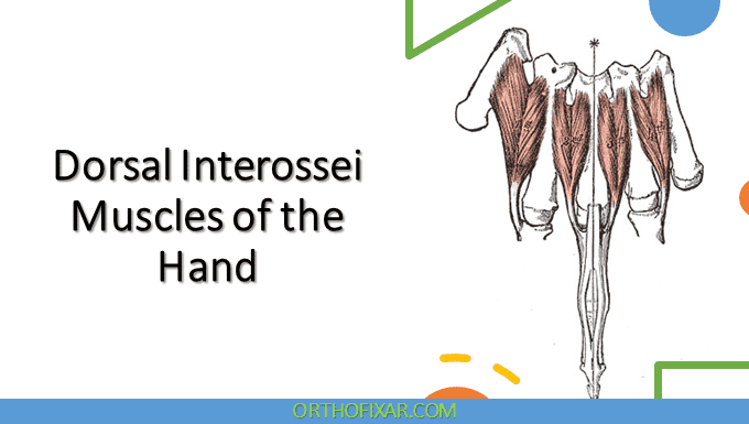
The four dorsal interosseous muscles (dorsal interossei) are located between the metacarpals; the 1st dorsal interosseous muscle is…
Read Full Entry →
The Opponens Digiti Minimi muscle originates from the hook of hamate and flexor retinaculum and inserts into the…
Read Full Entry →