
Elbow Ultrasound Imaging
The elbow joint presents an ideal anatomical region for ultrasound examination due to the relatively superficial location of…
Read Full Entry →
The elbow joint presents an ideal anatomical region for ultrasound examination due to the relatively superficial location of…
Read Full Entry →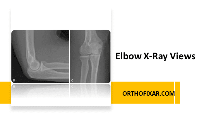
Plain radiographs remain the first-line imaging modality for evaluating elbow pathology. Elbow X-ray views provide essential information about…
Read Full Entry →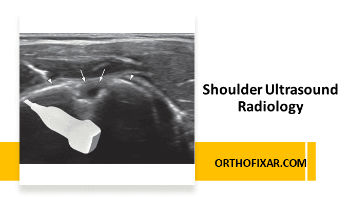
Shoulder ultrasound imaging modality allows for real-time, dynamic evaluation of shoulder structures and provides excellent visualization of soft…
Read Full Entry →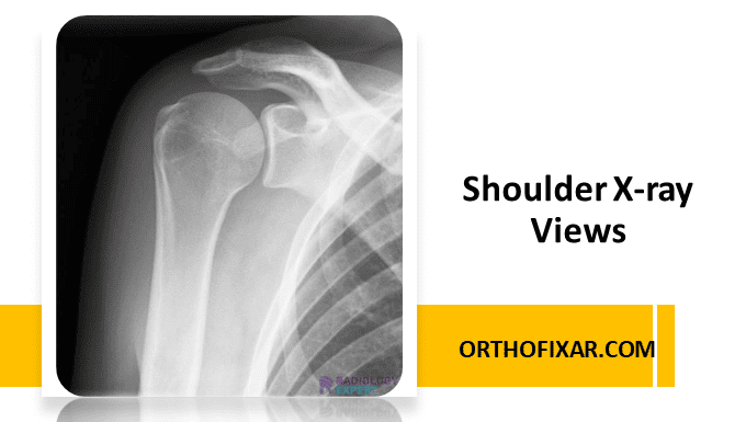
Diagnostic imaging is used in conjunction with a physical examination to determine a diagnosis. It should never be…
Read Full Entry →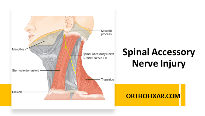
The spinal accessory nerve (cranial nerve XI) is a motor nerve that innervates the sternocleidomastoid and trapezius muscles.…
Read Full Entry →
The Gluteus Maximus Muscle has a wide origin, from posterior aspect of dorsal ilium, posterior to posterior gluteal…
Read Full Entry →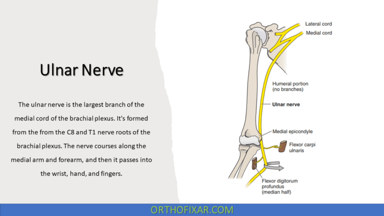
The ulnar nerve is the largest branch of the medial cord of the brachial plexus. It's formed from…
Read Full Entry →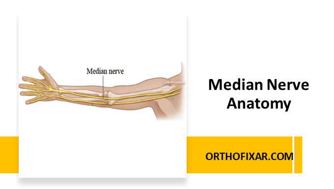
The median nerve arises from the lateral and medial cords of the brachial plexus, with contributions from spinal…
Read Full Entry →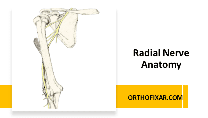
The radial nerve exits the axilla posterior to the brachial artery and enter the posterior arm compartment through…
Read Full Entry →
The axillary nerve arises from the posterior cord of the brachial plexus, carrying nerve fibers from the C5…
Read Full Entry →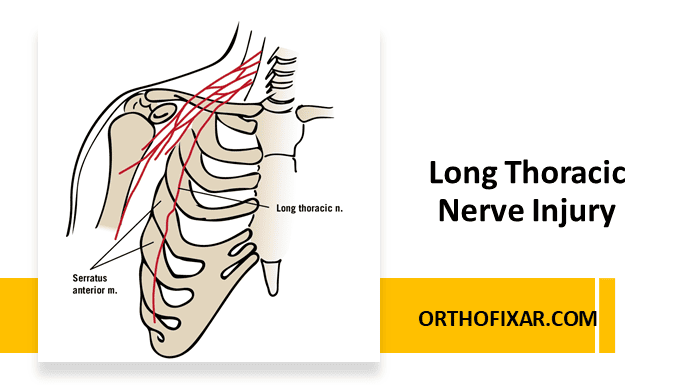
Long Thoracic Nerve Injury is an uncommon but significant etiology for scapular dysfunction. This nerve is mainly derived…
Read Full Entry →
After originating from the lateral cord, the musculocutaneous nerve pierces the coracobrachialis muscle approximately 5–8 cm distal to…
Read Full Entry →