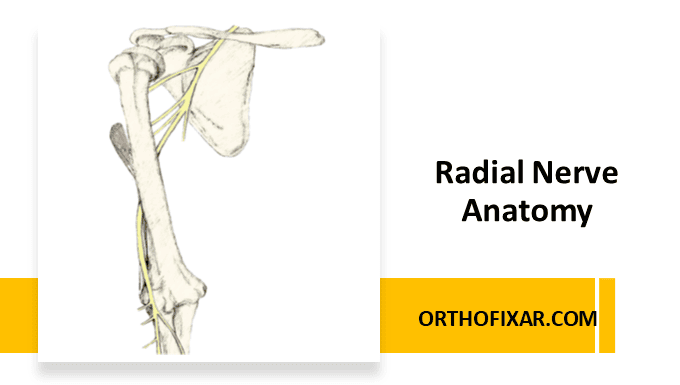
The radial nerve is the largest nerve arising from the posterior cord of the brachial plexus (C5-T1). It has both motor and sensory functions.
Radial Nerve Course
Shoulder Region
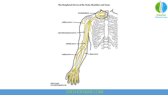
The radial nerve exits the axilla posterior to the brachial artery and enter the posterior arm compartment through the triangular interval, this space is bounded by the teres major muscle superiorly, the long head of the triceps muscle medially, and the lateral head of the triceps muscle laterally. In this location, the nerve is vulnerable to compression or injury during surgical procedures involving the posterior shoulder.
Humeral Course
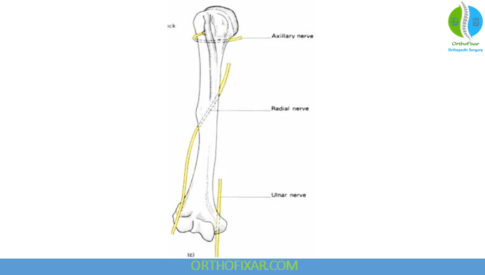
Spiral Groove Trajectory: The radial nerve spirals around the posterior aspect of the humeral shaft in the spiral groove (also known as the radial groove), traveling from medial to lateral.
Key anatomical landmarks include:
- Position approximately 20 cm proximal to the medial epicondyle
- Position approximately 14 cm proximal to the lateral epicondyle
These landmarks are important during surgical approaches to the humerus to dispose the radial nerve safely.
Lateral Emergence: After coursing through the spiral groove, the nerve pierces the lateral intermuscular septum and emerges on the lateral aspect of the arm approximately 7.5 cm above the trochlea. At this point, it lies in the interval between the brachialis muscle posteriorly and the brachioradialis muscle anteriorly.
Epicondylar Passage: The nerve passes anterior to the lateral epicondyle while maintaining its position between the brachialis and brachioradialis muscles. This anatomical relationship is crucial during lateral epicondylar surgical approaches.
Terminal Division
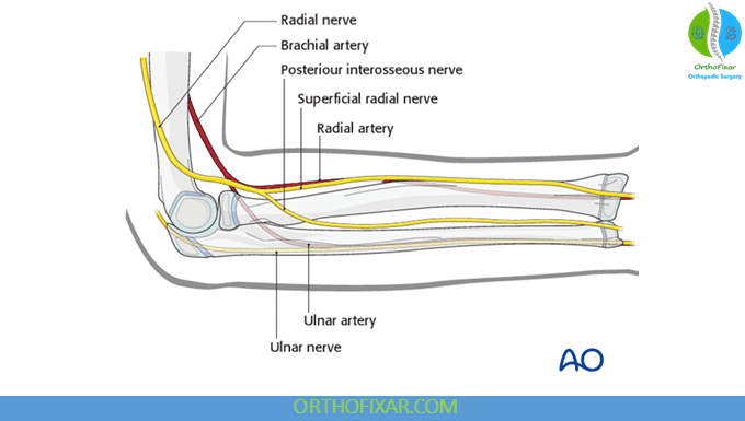
Approximately 1-3 cm distal to the lateral epicondyle, the radial nerve divides into its two terminal branches:
- Superficial branch (sensory)
- Deep branch (motor), also known as the posterior interosseous nerve (PIN)
Forearm Course
Deep Branch (Posterior Interosseous Nerve): This motor branch passes between the two heads of the supinator muscle, making it vulnerable to compression at this anatomical bottleneck. The nerve then travels along the posterior aspect of the interosseous membrane, innervating the deep extensor muscles of the forearm.
See Also: Radial Nerve Entrapment
Superficial Branch: This sensory branch follows the radial artery along the anterolateral aspect of the radius, traveling deep to the brachioradialis muscle. In the distal forearm, it courses dorsally over the distal radius, passing through the anatomical snuffbox to reach its cutaneous distribution.
Motor Innervation
Main Radial Nerve Trunk
The radial nerve innervates muscles in the arm before its terminal division:
Triceps Brachii: Both the medial and lateral heads receive innervation from the main trunk. The long head is typically innervated by a separate branch that arises more proximally.
Brachioradialis: This muscle receives innervation from the main radial nerve trunk, making it useful for clinical testing of radial nerve function proximal to the elbow.
Extensor Carpi Radialis Longus: Innervated by the main trunk, this muscle contributes to wrist extension and radial deviation.
Anconeus: This small muscle assists with elbow extension and receives innervation from the main radial nerve.
Deep Branch (Posterior Interosseous Nerve)
The deep branch provides motor innervation to the remaining extensor muscles of the forearm:
Proximal Muscles:
Distal Muscles:
- Extensor digitorum
- Extensor digiti minimi
- Extensor carpi ulnaris
- Abductor pollicis longus
- Extensor pollicis longus
- Extensor pollicis brevis
- Extensor indicis
Sensory Innervation
The radial nerve provides cutaneous sensation through several branches that arise at different levels along its course.
Anterior Aspect
Inferior Lateral Cutaneous Nerve of the Arm: This branch provides sensation to the anterior lateral aspect of the mid-arm, typically arising from the radial nerve in the spiral groove.
Posterior Aspect
Posterior Cutaneous Nerve of the Arm: Arising proximally, this branch innervates the posterior aspect of the distal arm.
Posterior Cutaneous Nerve of the Forearm: This branch provides sensation to a longitudinal strip along the posterior aspect of the forearm, extending from the elbow to the wrist.
Superficial Branch: The terminal sensory branch of the radial nerve provides sensation to:
- Posterior (dorsal) aspect of the thumb
- Posterior aspect of the index finger
- Posterior aspect of the middle finger
- Lateral half of the posterior aspect of the ring finger
- Associated dorsal hand area overlying these digits
Clinical Branches
Understanding the branching pattern of the radial nerve is essential for localizing injuries and planning surgical approaches:
- Posterior Cutaneous Nerve of the Arm
- Posterior Cutaneous Nerve of the Forearm
- Superficial Branch (terminal sensory branch)
- Posterior Interosseous Nerve (terminal motor branch)
Clinical Correlations
Humeral Fractures: The intimate relationship between the radial nerve and the humeral shaft makes it vulnerable to injury in mid-shaft humeral fractures, with reported incidence rates of 2-18%.
Posterior Interosseous Nerve Syndrome: Compression of the deep branch as it passes through the supinator muscle can result in motor weakness without sensory loss.
Superficial Branch Injury: Isolated injury to the superficial branch results in sensory loss over the anatomical snuffbox and dorsal hand, often described as numbness in the first web space.
Surgical Considerations: Knowledge of the radial nerve’s anatomical course is crucial during approaches to the humerus, lateral epicondyle, and posterior forearm to avoid iatrogenic injury.
References & More
- Glover NM, Black AC, Murphy PB. Anatomy, Shoulder and Upper Limb, Radial Nerve. [Updated 2023 Nov 5]. In: StatPearls [Internet]. Treasure Island (FL): StatPearls Publishing; 2025 Jan-. Available from: https://www.ncbi.nlm.nih.gov/books/NBK534840/
