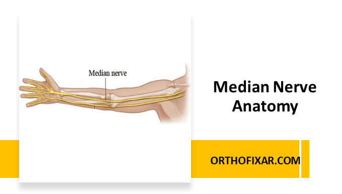
The median nerve arises from the lateral and medial cords of the brachial plexus, with contributions from spinal roots C6, C7, C8, and T1. It runs through the anterior portion of the arm and forearm, providing motor innervation to most of the flexor muscles in the forearm, as well as the thenar and lumbrical muscles in the hand. Additionally, it supplies sensory innervation to parts of the skin of the hand.
Median Nerve Anatomical Course
Shoulder and Arm Region
The median nerve accompanies the brachial artery throughout its course in the arm. The nerve crosses the brachial artery during its descent at approximately 15 centimeters proximal to the medial epicondyle where the nerve transitions from a lateral to medial position relative to the artery.
At the distal arm, the median nerve position is medial to the brachial artery and lies superficial to the brachialis muscle. The median nerve gives off no branches in the arm, distinguishing it from other major nerves of the upper extremity.

Forearm Region
The median nerve encounters the pronator teres muscle, where it characteristically splits the muscle’s two head, in this location the pronator syndrome may occur.
The median nerve then travels between the flexor digitorum superficialis (FDS) and flexor digitorum profundus (FDP) muscles.


Major Branch: Anterior Interosseous Nerve
The anterior interosseous nerve is a motor nerve branches that arises from the main trunk approximately 4 centimeters distal to the elbow joint. It then descends along the interosseous membrane, running between the flexor pollicis longus (FPL) and flexor digitorum profundus muscles.
The anterior interosseous nerve terminates in the pronator quadratus muscle, providing motor innervation to this deep compartment muscle. Injury to this nerve results in the characteristic “anterior interosseous syndrome,” which presents with weakness in thumb flexion and index finger flexion.
Wrist and Carpal Tunnel
As the median nerve approaches the wrist, it gives rise to the palmar cutaneous branch approximately 6 centimeters proximal to the radial styloid process. This branch is clinically significant because it passes superficial to the flexor retinaculum, avoiding the carpal tunnel entirely. This anatomical arrangement explains why sensation over the thenar eminence is typically preserved in carpal tunnel syndrome.
The main median nerve trunk passes through the carpal tunnel, positioned between the tendons of the flexor digitorum superficialis and the flexor carpi radialis. Nerve compression in this carpal tunnel leads to the common clinical condition known as carpal tunnel syndrome.
See Also: Carpal Tunnel Syndrome

Motor Innervation Patterns
The median nerve provides motor innervation to several muscles that are essential for hand function:
- Pronator teres: Enables forearm pronation
- Flexor carpi radialis: Contributes to wrist flexion and radial deviation
- Palmaris longus: Assists in wrist flexion (when present)
- Flexor digitorum superficialis: Provides superficial finger flexion
Hand muscles:
- Abductor pollicis brevis: Primary thumb abductor
- Superficial head of flexor pollicis brevis: Contributes to thumb flexion
- Opponens pollicis: Essential for thumb opposition
- First and second lumbrical muscles: Facilitate finger extension at interphalangeal joints
Anterior Interosseous Nerve
This branch supplies the deep flexor compartment muscles:
- Flexor digitorum profundus (radial half): Provides deep flexion to index and middle fingers
- Flexor pollicis longus: Enables thumb flexion at interphalangeal joint
- Pronator quadratus: Primary deep forearm pronator
Sensory Distribution
The median nerve provides sensory innervation to the volar (palmar) aspect of the thumb, index finger, middle finger, and the radial half of the ring finger.
The palmar cutaneous branch specifically innervates the skin over the thenar eminence, which, as mentioned previously, is typically spared in carpal tunnel syndrome due to its superficial course over the flexor retinaculum.

References & More
- Murphy KA, Morrisonponce D. Anatomy, Shoulder and Upper Limb, Median Nerve. [Updated 2023 Aug 20]. In: StatPearls [Internet]. Treasure Island (FL): StatPearls Publishing; 2025 Jan-. Available from: https://www.ncbi.nlm.nih.gov/books/NBK448084/
