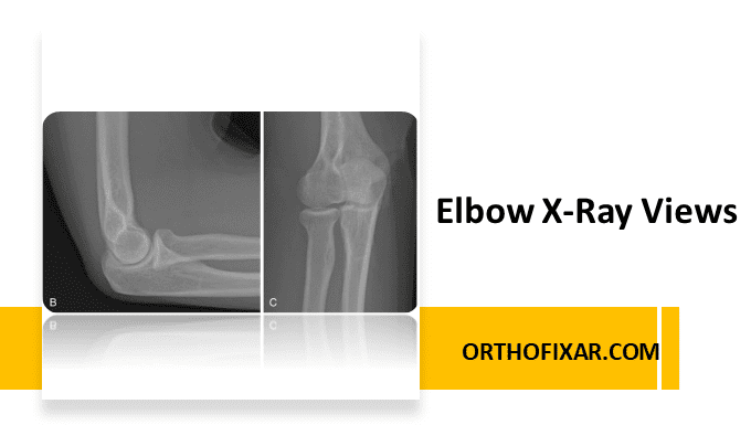
Plain radiographs remain the first-line imaging modality for evaluating elbow pathology. Elbow X-ray views provide essential information about bone alignment, joint spaces, and soft tissue changes. Selecting the appropriate elbow X-ray views depends on the clinical question—such as trauma assessment, evaluation for arthritis, or suspicion of instability.
Common Elbow X-Ray Views
Depending on the suspected pathology, the following X-ray views are commonly obtained:
- Anteroposterior (AP) view
- Lateral view at 90° flexion
- Cubital tunnel view
- Anteroposterior internal oblique view (often used in trauma)
- Anteroposterior external oblique view (often used in trauma)
- Axial view (for certain bony lesions and loose bodies)
See Also: Shoulder X-ray Views
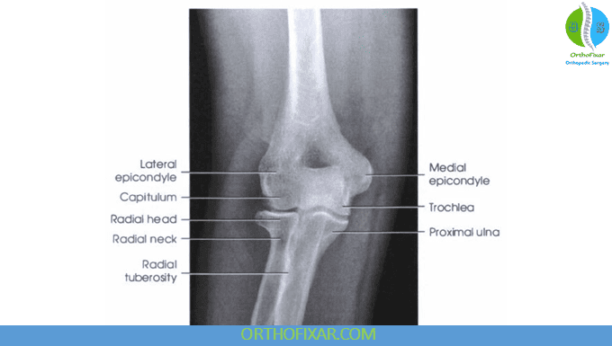
1. Elbow Anteroposterior (AP) View
Purpose: Provides a frontal view of the elbow joint, visualizing the distal humerus, proximal radius, and ulna.
Key points to assess:
- Bony landmarks: Epicondyles, trochlea, capitulum, radial head, radial tuberosity, coronoid process, and olecranon process.
- Pathology detection:
- Bone spurs, osteochondral lesions, enthesopathy
- Loose bodies, calcifications, myositis ossificans
- Joint space narrowing, osteophytes
- Instability signs: Slight widening of the ulnohumeral joint (“drop sign”) or posterior displacement of the radial head relative to the capitellum → may suggest posterolateral rotary instability.
- Pediatric consideration: Check epiphyseal plates for normal development—growth in the humerus occurs mainly at the shoulder, while the radius and ulna grow mostly at the wrist.
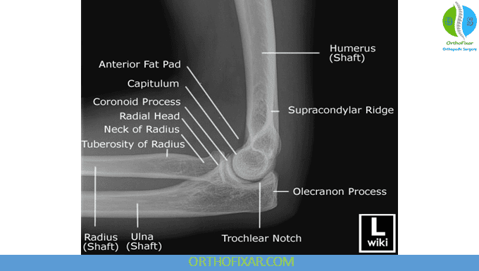
2. Elbow Lateral View (Elbow Flexed at 90°)
Purpose: Complements the AP view by providing a side profile of the elbow, useful for evaluating alignment and soft tissue signs.
Key points to assess:
- Same bony landmarks as in AP view.
- Soft tissue indicators:
- Fat-pad sign → suggests joint effusion; common in fractures, inflammatory arthritis, infection, or osteoid osteoma.
- Instability/trauma clues:
- Dislocations (posterior most common)
- Radial head dislocation may cause an impaction defect of the capitellum (Hill-Sachs–type lesion of the elbow).
- Can be used to measure the carrying angle.
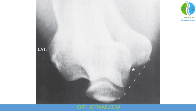
3. Cubital Tunnel View
Purpose: Specialized view to evaluate the cubital tunnel region, particularly in suspected ulnar nerve entrapment or bony narrowing.
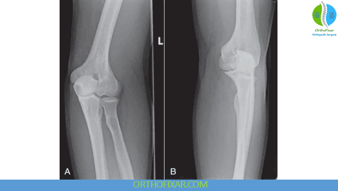
4. Elbow Oblique Views (Internal and External)
- Internal oblique: Helpful in trauma to visualize the radial head and coronoid process without overlap.
- External oblique: Used to assess lateral epicondyle and capitulum more clearly.
Practical Tips for Interpretation
- Always compare AP and lateral views—many injuries are missed on a single projection.
- Look for subtle alignment changes—even small displacements can indicate significant ligament injury.
- Check soft tissue shadows—especially anterior and posterior fat pads.
- In pediatrics, use an age-appropriate ossification chart to avoid mistaking normal growth centers for fractures.
References & More
- Goldfarb CA, Patterson JM, Sutter M, Krauss M, Steffen JA, Galatz L. Elbow radiographic anatomy: measurement techniques and normative data. J Shoulder Elbow Surg. 2012 Sep;21(9):1236-46. doi: 10.1016/j.jse.2011.10.026. Epub 2012 Feb 12. PMID: 22329911; PMCID: PMC3355210. PubMed
- Kane SF, Lynch JH, Taylor JC. Evaluation of elbow pain in adults. Am Fam Physician. 2014;89(8):649–657. PubMed
- Orthopedic Physical Assessment by David J. Magee, 7th Edition.


Closed.