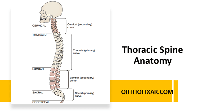
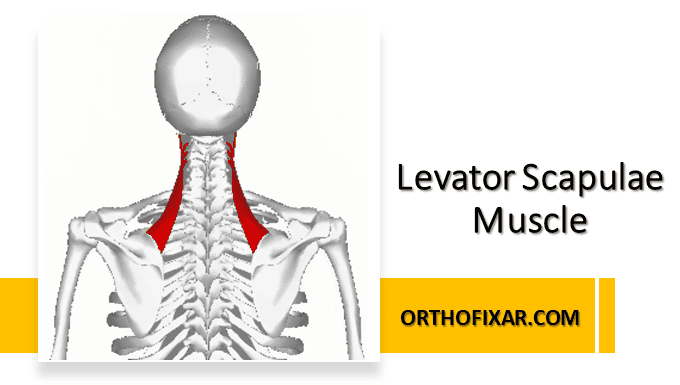
The thoracic spine consists of 12 vertebrae (T1-T12) that form the middle segment of the vertebral column. What distinguishes the thoracic spine from other spinal regions is its intimate relationship with the rib cage, creating a complex system of articulations that provide both stability and controlled mobility.
Thoracic Spine Vertebral Anatomy
The thoracic vertebrae exhibit unique size variations throughout the region. Interestingly, vertebrae diminish in size from T1 thoracic vertebrae to T3, then progressively increase in size toward T12 vertebrae.
The defining characteristic of thoracic vertebrae is the presence of facets on both the vertebral bodies and transverse processes for rib articulation.
Spinous Process Orientation
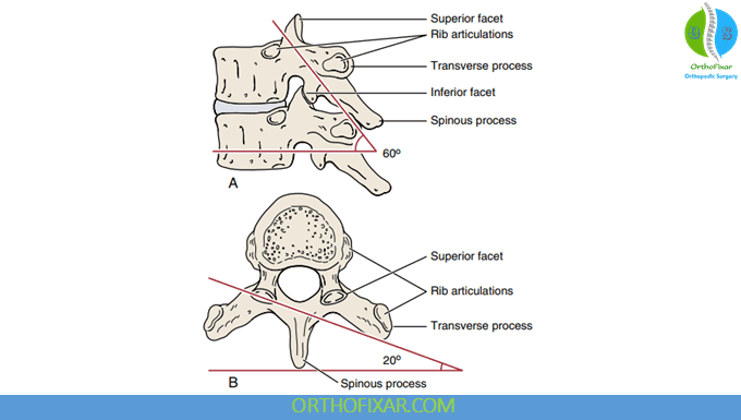
The spinous processes of thoracic spine vertebrae display a distinctive oblique downward orientation, with T7 vertebrae showing the greatest angulation.
The upper three thoracic vertebrae (T1-T3) have spinous processes that project directly posteriorly, positioning them on the same plane as their corresponding transverse processes. T4-T6 vertebrae show spinous processes that project slightly downward, with their tips positioned halfway between their own transverse processes and those of the vertebra below.
The maximum downward projection occurs at T7-T9, where the spinous process tips align with the transverse processes of the vertebra below. This relationship has important clinical implications – when applying posteroanterior pressure to the T8 spinous process, for example, you’re actually influencing the T9 vertebral body, while T8’s body may arc backward slightly.
T10 maintains the T9 pattern, while T11 returns to the T6 arrangement (halfway between transverse processes), and T12 vertebrae resembles T3 (level with its own transverse process).
See Also: Postural Alignment Assessment
Transitional Vertebrae
Several thoracic spine’s vertebrae are classified as transitional due to their unique characteristics. T1 thoracic vertebrae serves as a transition between cervical and thoracic regions, with its superior facet resembling cervical facets – facing upward and backward. T7 vertebrae is sometimes considered transitional as it represents the point where upper and lower limb axial rotation patterns alternate. T11 and T12 vertebrae are definitively transitional, with their facets becoming more similar to lumbar facets in orientation.
Facet Joint
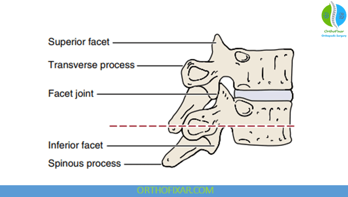
The facet joints (zygapophyseal joints) in the thoracic spine form the posterior elements of the classic “three-joint complex” along with the intervertebral disc. Their orientation changes systematically throughout the thoracic region, directly influencing movement patterns.
From T2-T11, the superior facets face upward, backward, and slightly laterally, while inferior facets face downward, forward, and slightly medially. The inclination angle progresses from 45-60° at T2-T3 to 90° at T4-T9. This configuration enables the slight rotation that characterizes thoracic spine movement while limiting flexion and anterior translation.
The transitional T11-T12 vertebrae adopt a more lumbar-like orientation, with superior facets facing upward, backward, and more medially, while inferior facets face forward and slightly laterally. The close-packed position is extension for all facet joints in the thoracic spine.
See Also: Spine Movements
Costovertebral Joint System
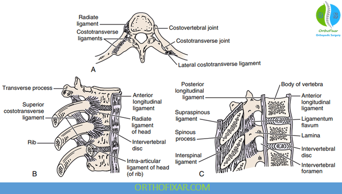
The connection between ribs and thoracic spine vertebrae occurs through two distinct joint systems: costovertebral joints and costotransverse joints.
Costovertebral Joints
These synovial plane joints exist at 24 locations between ribs and vertebral bodies. The system displays an elegant asymmetry: ribs 1, 10, 11, and 12 articulate with a single vertebra, while ribs 2-9 span two adjacent vertebrae and the intervening disc.
For the dual-vertebra articulations (ribs 2-9), an intraarticular ligament divides the joint into two compartments, allowing each rib head to contact both adjacent vertebral bodies and the intervertebral disc between them.
The primary stabilizing structure is the radiate ligament, which spreads from the anterior rib head like a fan to attach to the sides of vertebral bodies and, for ribs 2-9, the intervening disc. For ribs 10-12, this ligament attaches only to the adjacent vertebral body, reflecting their single-vertebra articulation pattern.
Costotransverse Joints
These synovial joints connect ribs 1-10 to the transverse processes at the same vertebral level of the thoracic spine. Notably, ribs 11 and 12 lack this articulation entirely, contributing to their classification as “floating ribs.”
Three ligaments support each costotransverse joint. The superior costotransverse ligament connects the transverse process above to the upper border of the rib and its neck. The costotransverse ligament proper runs between the rib neck and the same-level transverse process. Finally, the lateral costotransverse ligament extends from the transverse process tip to the adjacent rib.
Rib Cage
The 12 pairs of ribs create a protective cage around vital thoracic organs while permitting respiratory motion. Their classification reflects their distal attachments and functional roles.
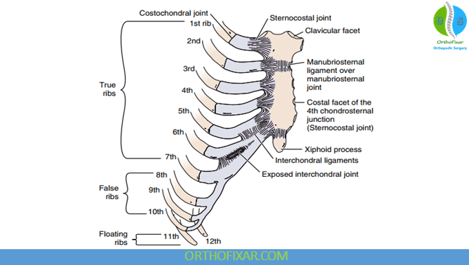
Ribs 1-7 are “true ribs,” articulating directly with the sternum through their costal cartilages. Ribs 8-10 are “false ribs,” connecting indirectly to the sternum by joining the costal cartilage of the rib above. Ribs 11-12 are “floating ribs,” with no anterior attachment to either sternum or costal cartilage.
The rib articulation pattern with vertebrae follows a systematic arrangement. The first rib articulates solely with T1 thoracic vertebrae, while ribs 2-9 each span two vertebrae (rib 2 contacts T1-T2, rib 3 contacts T2-T3, and so forth). Ribs 10-12 return to single-vertebra articulation with their corresponding vertebrae.
Anterior Chest Wall Joints
The costochondral joints connect ribs to their costal cartilages, while sternocostal joints link costal cartilages to the sternum. The first sternocostal joint is a synchondrosis (cartilaginous joint), while joints 2-6 are synovial. Where adjacent ribs or costal cartilages meet (ribs 5-9), synovial interchondral joints form.
Respiratory Biomechanics
Ribs transition from relatively horizontal positioning at the top of the cage to increasingly oblique orientations inferiorly, with the 12th rib being more vertical than horizontal.
During inspiration, three distinct movement patterns increase thoracic dimensions. The first six ribs primarily perform “pump-handle” motion, rotating around their long axes to increase anteroposterior diameter. This rotation involves upward neck movement during elevation and downward movement during depression, accompanied by forward and upward manubrium movement.
Ribs 7-10 execute “bucket-handle” motion, moving upward, backward, and medially during inspiration to increase the lateral (transverse) dimension and widen the infrasternal angle. Ribs 2-6 also contribute to this action but to a lesser degree.
The lower ribs (8-12) perform “caliper action,” moving laterally to increase lateral chest diameter. These coordinated movements efficiently expand the thoracic cavity for respiratory function.
Spinal Ligaments
The thoracic spine shares the standard complement of spinal ligaments found throughout the vertebral column. The ligamentum flavum connects adjacent laminae, while the anterior and posterior longitudinal ligaments run along the vertebral body surfaces. The interspinous and supraspinous ligaments connect adjacent spinous processes, and intertransverse ligaments span between transverse processes.
Clinical Correlations
The thoracic spine’s unique anatomy creates specific clinical considerations. The rib cage significantly stiffens this spinal region compared to cervical and lumbar areas, limiting mobility but providing stability. Age-related changes affect rib elasticity, with ribs becoming increasingly brittle over time, influencing fracture patterns and healing considerations.
The relationship between spinous process position and vertebral body location becomes crucial during manual examination and treatment. Understanding that pressure applied to a spinous process may primarily affect the vertebral body below guides precise therapeutic interventions.
The transitional nature of certain vertebrae (T1, T7, T11-T12 vertebrae) creates regions where movement patterns and biomechanical stresses change, often making these areas more susceptible to dysfunction or injury.
See Also: Spine Examination
References & More
- Williams P, Warwick R, eds. Gray’s Anatomy. 36th ed. British, Edinburgh: Churchill Livingstone; 1980.
- Waxenbaum JA, Reddy V, Futterman B. Anatomy, Back, Thoracic Vertebrae. [Updated 2023 Aug 1]. In: StatPearls [Internet]. Treasure Island (FL): StatPearls Publishing; 2025 Jan-. Available from: PubMed
- Orthopedic Physical Assessment by David J. Magee, 7th Edition.
